Tuesday Poster Session
Category: General Endoscopy
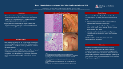
P4141 - From Polyp to Pathogen: Atypical MAC Infection Presentation on EGD
Tuesday, October 29, 2024
10:30 AM - 4:00 PM ET
Location: Exhibit Hall E

Has Audio
.jpg)
Sabrina Ho, MD
University of Arizona College of Medicine
Tucson, AZ
Presenting Author(s)
Brandon Witten, MD1, Sabrina Ho, MD1, Samuel H. Cheong, DO2, Jamie Scott, MD1, Sara Fallahi, MD1, Cameron M. Thompson, MD1
1University of Arizona College of Medicine, Tucson, AZ; 2Banner - University of Arizona, Tucson, AZ
Introduction: GI involvement from atypical organisms in severely immunocompromised hosts is a well known phenomenon. CMV, giardia, cryptosporidium are all organisms that are frequently encountered in AIDs patients. Non-tuberculosis mycobacterium avium complex (MAC) infections are a rare cause of GI manifestations of severely low CD4 counts.
Case Description/Methods: 46 year old male past medical history significant for HIV with resultant AIDs with consistently low CD4 counts despite antiretroviral therapy. Presented for evaluation for approximately 2.5 months of ongoing/worsening abdominal distention and watery diarrhea. Abdominal CT revealed fluid filled loops of small bowel as well as bulky mesenteric/retroperitoneal lymphadenopathy. Additionally, there was evidence of chronic liver disease and portal hypertension, splenomegaly and moderate volume ascites. GI was consulted for clinically significant diarrhea of unknown origin in the setting of an immunocompromised host. Significant findings during colonoscopy were four small polyps in the cecum as well as pan-colonic friability of which random colonic biopsies were taken. Pathology revealed abundant acid fast bacilli present forming nodules in the cecum as well as throughout the entire colon. EGD showed large atypical looking nodules within the duodenum which also had abundant acid fast bacilli. Unfortunately the patient later passed away from complications due to cirrhosis.
Discussion: Non-tuberculosis MAC infection has been described endoscopically as small miliary like lesions frequently encountered in patients who are severely immunocompromised. In the literature there are rare descriptions of mass like lesions caused by MAC infections, however, ours is one of the first to describe bulky lesions present endoscopically in the small and large intestine respectively.

Disclosures:
Brandon Witten, MD1, Sabrina Ho, MD1, Samuel H. Cheong, DO2, Jamie Scott, MD1, Sara Fallahi, MD1, Cameron M. Thompson, MD1. P4141 - From Polyp to Pathogen: Atypical MAC Infection Presentation on EGD, ACG 2024 Annual Scientific Meeting Abstracts. Philadelphia, PA: American College of Gastroenterology.
1University of Arizona College of Medicine, Tucson, AZ; 2Banner - University of Arizona, Tucson, AZ
Introduction: GI involvement from atypical organisms in severely immunocompromised hosts is a well known phenomenon. CMV, giardia, cryptosporidium are all organisms that are frequently encountered in AIDs patients. Non-tuberculosis mycobacterium avium complex (MAC) infections are a rare cause of GI manifestations of severely low CD4 counts.
Case Description/Methods: 46 year old male past medical history significant for HIV with resultant AIDs with consistently low CD4 counts despite antiretroviral therapy. Presented for evaluation for approximately 2.5 months of ongoing/worsening abdominal distention and watery diarrhea. Abdominal CT revealed fluid filled loops of small bowel as well as bulky mesenteric/retroperitoneal lymphadenopathy. Additionally, there was evidence of chronic liver disease and portal hypertension, splenomegaly and moderate volume ascites. GI was consulted for clinically significant diarrhea of unknown origin in the setting of an immunocompromised host. Significant findings during colonoscopy were four small polyps in the cecum as well as pan-colonic friability of which random colonic biopsies were taken. Pathology revealed abundant acid fast bacilli present forming nodules in the cecum as well as throughout the entire colon. EGD showed large atypical looking nodules within the duodenum which also had abundant acid fast bacilli. Unfortunately the patient later passed away from complications due to cirrhosis.
Discussion: Non-tuberculosis MAC infection has been described endoscopically as small miliary like lesions frequently encountered in patients who are severely immunocompromised. In the literature there are rare descriptions of mass like lesions caused by MAC infections, however, ours is one of the first to describe bulky lesions present endoscopically in the small and large intestine respectively.

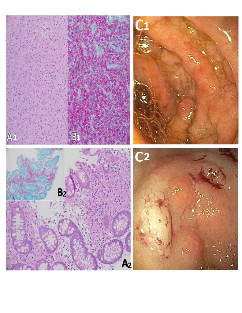
Figure: A1: Duodenal submucosal nodule with numerous foamy macrophages
B1: AFB stained nodule demonstrating substantial acid-fast bacilli in macrophages
C1: Endoscopic view of duodenal polypoid lesions representing severe MAC infection (A1/B1 source)
A2: Colonic biopsy demonstrating colonic mucosa with numerous foamy macrophages
B2: Colonic biopsy demonstrating macrophages filled with abundant acid-fast bacilli
C2: Endoscopic view of cecal polyp representing severe MAC infection (A2/B2 source)
Disclosures:
Brandon Witten indicated no relevant financial relationships.
Sabrina Ho indicated no relevant financial relationships.
Samuel Cheong indicated no relevant financial relationships.
Jamie Scott indicated no relevant financial relationships.
Sara Fallahi indicated no relevant financial relationships.
Cameron Thompson indicated no relevant financial relationships.
Brandon Witten, MD1, Sabrina Ho, MD1, Samuel H. Cheong, DO2, Jamie Scott, MD1, Sara Fallahi, MD1, Cameron M. Thompson, MD1. P4141 - From Polyp to Pathogen: Atypical MAC Infection Presentation on EGD, ACG 2024 Annual Scientific Meeting Abstracts. Philadelphia, PA: American College of Gastroenterology.
