Tuesday Poster Session
Category: Esophagus
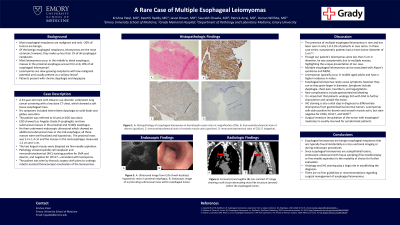
P3978 - A Rare Case of Multiple Esophageal Leiomyomas
Tuesday, October 29, 2024
10:30 AM - 4:00 PM ET
Location: Exhibit Hall E

Has Audio

Krishna Patel, MD
Emory University School of Medicine
Atlanta, GA
Presenting Author(s)
Krishna Patel, MD, Keerthi Reddy, MD, Jason Brown, MD, Saurabh Chawla, MD, Patrick Arraj, MD, Dorian Willhite, MD
Emory University School of Medicine, Atlanta, GA
Introduction: Leiomyomas are the most common type of benign esophageal neoplasm; however, they are rare and make up less than 1% of all esophageal neoplasms. Most leiomyomas occur in the middle to distal esophagus; masses in the proximal esophagus account for only 10% of all esophageal leiomyomas. Leiomyomas are slow-growing neoplasms with low malignant potential and usually present as a solitary lesion. Patients present with chronic dysphagia and dyspepsia. We present a unique case of a male with dysphagia found to have multiple esophageal leiomyomas.
Case Description/Methods: A 63-year-old male with tobacco use disorder underwent lung cancer screening with a low dose CT chest, which showed a soft tissue esophageal mass. The patient was referred to gastroenterology. EGD showed Los Angeles Grade D esophagitis and two submucosal masses in the proximal and middle esophagus. His symptoms included intermittent dysphagia to solid foods and globus sensation. He then underwent endoscopic ultrasound (EUS) which showed an additional submucosal mass in the mid-esophagus; all three masses were well-localized and hypoechoic. The proximal mass was 2.4 x 1.4 cm and the masses in the mid-esophagus measured 1.2 cm and 1 cm. The two largest masses were biopsied via fine-needle aspiration (FNA). Pathology showed spindle cell neoplasm and immunohistochemical (IHC) staining positive for SMA and desmin, and negative for CD117—consistent with leiomyoma. The patient was seen by thoracic surgery with plans to undergo robotic-assisted thoracoscopic enucleation of the leiomyomas.
Discussion: The presence of multiple esophageal leiomyomas is rare and has been seen in only 1.8-2.5% of patients in case series. In these case series, symptomatic patients had a mean tumor diameter of 5 cm1-2. Though our patient’s leiomyomas were less than 5 cm in diameter, he was symptomatic due to multiple masses, highlighting the unique presentation of our case. It is important that patients undergo EUS with FNA to further characterize and sample the lesion. IHC staining is also a vital step in diagnosis to differentiate leiomyomas from gastrointestinal stromal tumors. Surgical resection is usually reserved for symptomatic patients.
1. Jiang W, Rice TW, Goldblum JR. Esophageal leiomyoma: experience from a single institution. Diseases of the Esophagus. 2013;26(2):167-74.
2.Mutrie CJ, Donahue DM, Wain JC, Wright CD, Gaissert HA, Grillo HC, Mathisen DJ, Allan JS. Esophageal leiomyoma: a 40-year experience. The Annals of thoracic surgery. 2005;79(4):1122-5.

Disclosures:
Krishna Patel, MD, Keerthi Reddy, MD, Jason Brown, MD, Saurabh Chawla, MD, Patrick Arraj, MD, Dorian Willhite, MD. P3978 - A Rare Case of Multiple Esophageal Leiomyomas, ACG 2024 Annual Scientific Meeting Abstracts. Philadelphia, PA: American College of Gastroenterology.
Emory University School of Medicine, Atlanta, GA
Introduction: Leiomyomas are the most common type of benign esophageal neoplasm; however, they are rare and make up less than 1% of all esophageal neoplasms. Most leiomyomas occur in the middle to distal esophagus; masses in the proximal esophagus account for only 10% of all esophageal leiomyomas. Leiomyomas are slow-growing neoplasms with low malignant potential and usually present as a solitary lesion. Patients present with chronic dysphagia and dyspepsia. We present a unique case of a male with dysphagia found to have multiple esophageal leiomyomas.
Case Description/Methods: A 63-year-old male with tobacco use disorder underwent lung cancer screening with a low dose CT chest, which showed a soft tissue esophageal mass. The patient was referred to gastroenterology. EGD showed Los Angeles Grade D esophagitis and two submucosal masses in the proximal and middle esophagus. His symptoms included intermittent dysphagia to solid foods and globus sensation. He then underwent endoscopic ultrasound (EUS) which showed an additional submucosal mass in the mid-esophagus; all three masses were well-localized and hypoechoic. The proximal mass was 2.4 x 1.4 cm and the masses in the mid-esophagus measured 1.2 cm and 1 cm. The two largest masses were biopsied via fine-needle aspiration (FNA). Pathology showed spindle cell neoplasm and immunohistochemical (IHC) staining positive for SMA and desmin, and negative for CD117—consistent with leiomyoma. The patient was seen by thoracic surgery with plans to undergo robotic-assisted thoracoscopic enucleation of the leiomyomas.
Discussion: The presence of multiple esophageal leiomyomas is rare and has been seen in only 1.8-2.5% of patients in case series. In these case series, symptomatic patients had a mean tumor diameter of 5 cm1-2. Though our patient’s leiomyomas were less than 5 cm in diameter, he was symptomatic due to multiple masses, highlighting the unique presentation of our case. It is important that patients undergo EUS with FNA to further characterize and sample the lesion. IHC staining is also a vital step in diagnosis to differentiate leiomyomas from gastrointestinal stromal tumors. Surgical resection is usually reserved for symptomatic patients.
1. Jiang W, Rice TW, Goldblum JR. Esophageal leiomyoma: experience from a single institution. Diseases of the Esophagus. 2013;26(2):167-74.
2.Mutrie CJ, Donahue DM, Wain JC, Wright CD, Gaissert HA, Grillo HC, Mathisen DJ, Allan JS. Esophageal leiomyoma: a 40-year experience. The Annals of thoracic surgery. 2005;79(4):1122-5.

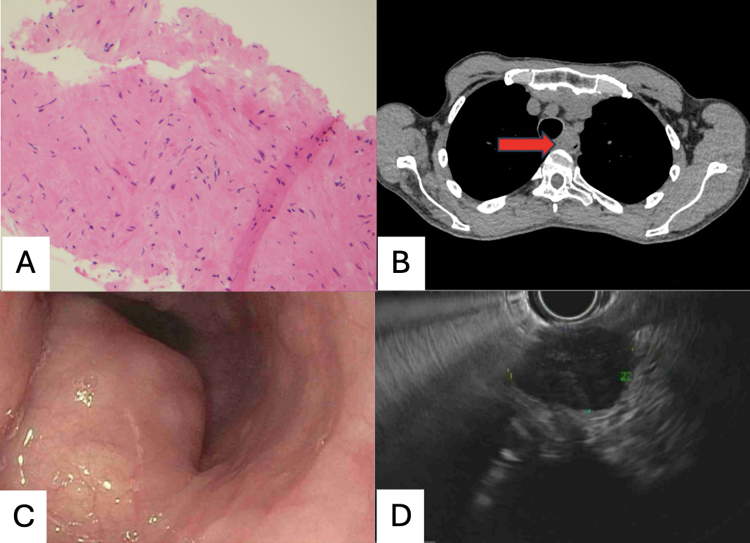
Figure: A: Histopathology of esophageal leiomyoma in hematoxylin-eosin stain at magnification 200x. B: Axial noncontrast CT image showing a soft tissue attenuating mass-like structure (arrow) within the esophageal lumen. C: Endoscopic image of a protruding submucosal mass within esophageal lumen. D: Ultrasound image from EUS of well-localized, hypoechoic mass in proximal esophagus.
Disclosures:
Krishna Patel indicated no relevant financial relationships.
Keerthi Reddy indicated no relevant financial relationships.
Jason Brown indicated no relevant financial relationships.
Saurabh Chawla indicated no relevant financial relationships.
Patrick Arraj indicated no relevant financial relationships.
Dorian Willhite indicated no relevant financial relationships.
Krishna Patel, MD, Keerthi Reddy, MD, Jason Brown, MD, Saurabh Chawla, MD, Patrick Arraj, MD, Dorian Willhite, MD. P3978 - A Rare Case of Multiple Esophageal Leiomyomas, ACG 2024 Annual Scientific Meeting Abstracts. Philadelphia, PA: American College of Gastroenterology.
