Tuesday Poster Session
Category: Esophagus
P3984 - Dual HSV/H. pylori Infection Mimicking an Esophagopleural Fistula: A Case Report
Tuesday, October 29, 2024
10:30 AM - 4:00 PM ET
Location: Exhibit Hall E

Has Audio

Anand Prabhu, MD
University of Illinois College of Medicine
Chicago, IL
Presenting Author(s)
Anand Prabhu, MD, Mary Biglin, MD
University of Illinois College of Medicine, Chicago, IL
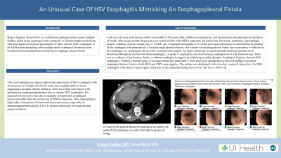
Introduction: Herpes Simplex Virus (HSV) esophagitis is a well-known pathogen which causes multiple shallow ulcers in the esophagus most commonly in immunosuppressed patients. It presents with odynophagia or chest pain and responds well to acyclovir or valacyclovir and complications rarely occur. This case depicts an atypical presentation of likely chronic HSV esophagitis in an AIDs patient presenting with multiple small esophageal diverticula with resultant pneumomediastinum mimicking an esophago-pleural fistula.
Case Description/Methods: A 48-year-old male with history of HIV on HAART (CD4 count 300), ESRD on hemodialysis, and hypertension was admitted for shortness of breath, after being recently diagnosed at an outside facility with MRSA empyema. He denied any chest pain, dysphagia, odynophagia, nausea, vomiting, melena, weight loss, or NSAID use. Computed tomography (CT) of the chest upon admission revealed diffuse thickening of the esophagus with pneumatosis, a loculated right pleural effusion, and concern for esophagopleural fistula due to proximity of effusion to the esophagus. An esophagram did not show contrast extravasation. An upper endoscopy revealed multiple small and medium sized diverticula throughout the mid and distal esophagus, LA grade C esophagitis, one distal oozing esophageal ulcer with blood oozing. There was no evidence of perforation, fistula, or dilated esophagus to suggest an underlying motility disorder. Esophageal biopsies showed HSV esophagitis. Notably, a limited report of an upper endoscopy performed 3 years prior at an outside facility showed multiple excavated esophageal lesions, however both HSV and CMV were negative. The patient was discharged with a ten-day course of valacyclovir for HSV esophagitis with plans to repeat upper endoscopy in the outpatient setting however he was lost to follow up.
Discussion: This case highlights an atypical endoscopic appearance of HSV esophagitis with the presence of multiple diverticula rather than multiple shallow ulcers, suggesting a possible chronic infection. There have been case reports with spontaneous esophageal perforation due to chronic HSV esophagitis. His pneumatosis may have been due to multiple asymptomatic esophageal diverticula rather than the red herring of MRSA empyema. Thus, maintaining a high index of suspicion of concurrent disease processes, especially in immunosuppressed patients, is key to prompt endoscopic investigation and proper treatment.
Disclosures:
Anand Prabhu, MD, Mary Biglin, MD. P3984 - Dual HSV/<i>H. pylori</i> Infection Mimicking an Esophagopleural Fistula: A Case Report, ACG 2024 Annual Scientific Meeting Abstracts. Philadelphia, PA: American College of Gastroenterology.
University of Illinois College of Medicine, Chicago, IL
Introduction: Herpes Simplex Virus (HSV) esophagitis is a well-known pathogen which causes multiple shallow ulcers in the esophagus most commonly in immunosuppressed patients. It presents with odynophagia or chest pain and responds well to acyclovir or valacyclovir and complications rarely occur. This case depicts an atypical presentation of likely chronic HSV esophagitis in an AIDs patient presenting with multiple small esophageal diverticula with resultant pneumomediastinum mimicking an esophago-pleural fistula.
Case Description/Methods: A 48-year-old male with history of HIV on HAART (CD4 count 300), ESRD on hemodialysis, and hypertension was admitted for shortness of breath, after being recently diagnosed at an outside facility with MRSA empyema. He denied any chest pain, dysphagia, odynophagia, nausea, vomiting, melena, weight loss, or NSAID use. Computed tomography (CT) of the chest upon admission revealed diffuse thickening of the esophagus with pneumatosis, a loculated right pleural effusion, and concern for esophagopleural fistula due to proximity of effusion to the esophagus. An esophagram did not show contrast extravasation. An upper endoscopy revealed multiple small and medium sized diverticula throughout the mid and distal esophagus, LA grade C esophagitis, one distal oozing esophageal ulcer with blood oozing. There was no evidence of perforation, fistula, or dilated esophagus to suggest an underlying motility disorder. Esophageal biopsies showed HSV esophagitis. Notably, a limited report of an upper endoscopy performed 3 years prior at an outside facility showed multiple excavated esophageal lesions, however both HSV and CMV were negative. The patient was discharged with a ten-day course of valacyclovir for HSV esophagitis with plans to repeat upper endoscopy in the outpatient setting however he was lost to follow up.
Discussion: This case highlights an atypical endoscopic appearance of HSV esophagitis with the presence of multiple diverticula rather than multiple shallow ulcers, suggesting a possible chronic infection. There have been case reports with spontaneous esophageal perforation due to chronic HSV esophagitis. His pneumatosis may have been due to multiple asymptomatic esophageal diverticula rather than the red herring of MRSA empyema. Thus, maintaining a high index of suspicion of concurrent disease processes, especially in immunosuppressed patients, is key to prompt endoscopic investigation and proper treatment.
Disclosures:
Anand Prabhu indicated no relevant financial relationships.
Mary Biglin indicated no relevant financial relationships.
Anand Prabhu, MD, Mary Biglin, MD. P3984 - Dual HSV/<i>H. pylori</i> Infection Mimicking an Esophagopleural Fistula: A Case Report, ACG 2024 Annual Scientific Meeting Abstracts. Philadelphia, PA: American College of Gastroenterology.
