Monday Poster Session
Category: Liver
P2997 - Elevated Alpha Fetoprotein of Unclear Origin: A New Subtype?
Monday, October 28, 2024
10:30 AM - 4:00 PM ET
Location: Exhibit Hall E

Has Audio
- NV
Nevin Varghese, MD
Thomas Jefferson University Hospital
Philadelphia, PA
Presenting Author(s)
Nevin Varghese, MD1, Madison Force, MD1, Alisha Goyal, BS2, Hie-Won Hann, MD1
1Thomas Jefferson University Hospital, Philadelphia, PA; 2Sidney Kimmel Medical College at Thomas Jefferson University, Philadelphia, PA
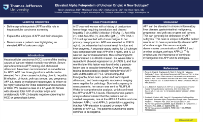
Introduction: Hepatocellular carcinoma (HCC) is one of the leading causes of cancer-related mortality worldwide. Serum alpha fetoprotein (AFP) testing and abdominal ultrasound have been recommended as surveillance for HCC. AFP has three glycoforms which can be elevated from other causes including chronic hepatitis B infection, cirrhosis, yolk sac tumors, and pregnancy. AFP-L3, made by malignant hepatocytes, is known to be highly sensitive for initial detection and recurrence of HCC. We present a case of a 67-year-old female with elevated total AFP of unclear origin and undetectable AFP-L3 despite negative screening for HCC or gynecologic tumor.
Case Description/Methods: A 67-year-old woman with a history of postpartum hemorrhage requiring transfusion and cleared hepatitis B virus (HBV) infection (HBsAg (-), Anti-HBs (+), total Anti-HBc (+), Anti-HBc IgM (-), HBV DNA < 10 IU/mL) presented with chronic fatigue to her primary care physician. AFP was elevated to 1292.9 ng/mL, but otherwise had normal renal function and liver enzymes. A separate assay testing for L3 subtype was completed with total AFP of 9.1 ng/mL and % L3 was too low for measurement. HCC screening MRI demonstrated a LI-RADS 4 lesion. Six weeks later a repeat MRI showed regression to LI-RADS 3, and four months later this lesion was found to be a pseudo-lesion due to vascular branching. Over the years, testing demonstrated a steadily rising level of AFP with undetectable AFP-L3. Chest computed tomography, bone scan, pelvic and transvaginal ultrasound, and brain magnetic resonance imaging were negative for pathology. Due to discrepancy in measurements, her serum was sent to FUJIFILM Wako for comprehensive analysis, which confirmed the AFP and AFP-L3 levels. Electrophoresis pattern analysis demonstrated that the patient’s serum created two peaks, one at the AFP-L1 fraction and one between AFP-L1 and AFP-L3, potentially suggesting that her AFP elevation is caused by a new AFP subtype or AFP-L2. The patient’s surveillance scans continue to be negative.
Discussion: AFP can be elevated in chronic inflammatory liver disease, gastrointestinal malignancy, pregnancy, and yolk sac or germ cell tumors. This can generally be delineated by AFP subtypes. This case is unique in that the patient was found to have a persistently elevated AFP of unclear origin. Her serum analysis demonstrates concentration of AFP-L1 and another subtype, perhaps AFP-L2. This emphasizes the importance of continued investigation into AFP and its etiologies.
Disclosures:
Nevin Varghese, MD1, Madison Force, MD1, Alisha Goyal, BS2, Hie-Won Hann, MD1. P2997 - Elevated Alpha Fetoprotein of Unclear Origin: A New Subtype?, ACG 2024 Annual Scientific Meeting Abstracts. Philadelphia, PA: American College of Gastroenterology.
1Thomas Jefferson University Hospital, Philadelphia, PA; 2Sidney Kimmel Medical College at Thomas Jefferson University, Philadelphia, PA
Introduction: Hepatocellular carcinoma (HCC) is one of the leading causes of cancer-related mortality worldwide. Serum alpha fetoprotein (AFP) testing and abdominal ultrasound have been recommended as surveillance for HCC. AFP has three glycoforms which can be elevated from other causes including chronic hepatitis B infection, cirrhosis, yolk sac tumors, and pregnancy. AFP-L3, made by malignant hepatocytes, is known to be highly sensitive for initial detection and recurrence of HCC. We present a case of a 67-year-old female with elevated total AFP of unclear origin and undetectable AFP-L3 despite negative screening for HCC or gynecologic tumor.
Case Description/Methods: A 67-year-old woman with a history of postpartum hemorrhage requiring transfusion and cleared hepatitis B virus (HBV) infection (HBsAg (-), Anti-HBs (+), total Anti-HBc (+), Anti-HBc IgM (-), HBV DNA < 10 IU/mL) presented with chronic fatigue to her primary care physician. AFP was elevated to 1292.9 ng/mL, but otherwise had normal renal function and liver enzymes. A separate assay testing for L3 subtype was completed with total AFP of 9.1 ng/mL and % L3 was too low for measurement. HCC screening MRI demonstrated a LI-RADS 4 lesion. Six weeks later a repeat MRI showed regression to LI-RADS 3, and four months later this lesion was found to be a pseudo-lesion due to vascular branching. Over the years, testing demonstrated a steadily rising level of AFP with undetectable AFP-L3. Chest computed tomography, bone scan, pelvic and transvaginal ultrasound, and brain magnetic resonance imaging were negative for pathology. Due to discrepancy in measurements, her serum was sent to FUJIFILM Wako for comprehensive analysis, which confirmed the AFP and AFP-L3 levels. Electrophoresis pattern analysis demonstrated that the patient’s serum created two peaks, one at the AFP-L1 fraction and one between AFP-L1 and AFP-L3, potentially suggesting that her AFP elevation is caused by a new AFP subtype or AFP-L2. The patient’s surveillance scans continue to be negative.
Discussion: AFP can be elevated in chronic inflammatory liver disease, gastrointestinal malignancy, pregnancy, and yolk sac or germ cell tumors. This can generally be delineated by AFP subtypes. This case is unique in that the patient was found to have a persistently elevated AFP of unclear origin. Her serum analysis demonstrates concentration of AFP-L1 and another subtype, perhaps AFP-L2. This emphasizes the importance of continued investigation into AFP and its etiologies.
Disclosures:
Nevin Varghese indicated no relevant financial relationships.
Madison Force indicated no relevant financial relationships.
Alisha Goyal indicated no relevant financial relationships.
Hie-Won Hann indicated no relevant financial relationships.
Nevin Varghese, MD1, Madison Force, MD1, Alisha Goyal, BS2, Hie-Won Hann, MD1. P2997 - Elevated Alpha Fetoprotein of Unclear Origin: A New Subtype?, ACG 2024 Annual Scientific Meeting Abstracts. Philadelphia, PA: American College of Gastroenterology.
