Monday Poster Session
Category: Liver
P3000 - Cyclical Right Upper Quadrant Pain: A Rare Presentation of Hepatic Endometriosis
Monday, October 28, 2024
10:30 AM - 4:00 PM ET
Location: Exhibit Hall E

Has Audio

Aditya Kler, MD
University of Minnesota
Minneapolis, MN
Presenting Author(s)
Award: Presidential Poster Award
Aditya Kler, MD1, Nirjhar Dutta, DO, MS2, Jennifer W. Haglund, MD3
1University of Minnesota, Minneapolis, MN; 2University of Minnesota Medical Center, Minneapolis, MN; 3Minneapolis VA Health Care System, Minneapolis, MN
Introduction: Endometriosis affects 10 % of women in their reproductive age. Here we present a rare presentation of endometriosis as a hepatic mass.
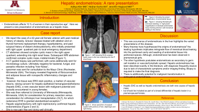
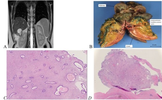
Case Description/Methods: We report the case of a 42-year-old female with past medical history of obesity, Graves’ disease treated with ablation and on thyroid hormone replacement therapy, hyperlipidemia, and past surgical history of distant cholecystectomy, who initially presented with right upper quadrant pain to local emergency department. CT abdomen showed complex cystic mass in the right upper quadrant, extending into right renal fossa. Subsequent MRI abdomen confirmed complex cystic mass (Figure 1A). A CT guided biopsy was performed, with cores additionally sent for microbiology culture, specimen was sent for consultation to a tertiary care center. The biopsy revealed fibroconnective and adipose tissue with inflammatory changes and fibrosis; however, tissue was ERG stain positive, a marker of vascular lesions, raising concern for hepatic epithelioid hemangioendothelioma (hepatic EHE), a rare vascular lesion with malignant potential. She was referred to a liver transplant center for consideration for primary resection versus orthotopic liver transplantation (OLT), for which extensive hepatic EHE is granted standardized exception. Hepatic segmentectomy with right nephrectomy confirmed hepatic endometriosis (Figure 1B,C,D). She is now pain free and doing well clinically.
Discussion: Only 28 known cases of isolated hepatic endometriosis have been reported in medical literature.
Many theories have hypothesized the origins of endometriosis: the leading hypothesis implicates retrograde flow of menstrual blood leaking into the peritoneal cavity and seeding of endometrial tissue into peritoneal spaces. Although, endometriosis has been observed in premenstrual girls. The other hypothesis postulates endometriosis as secondary to germ cell mutation or vascular/lymphatic spread. Diagnosis is frequently elusive due to lack of characteristic symptoms and/or imaging finding. Biopsy is required to make the final diagnosis. There is additionally potential for malignant transformation in extra-pelvic endometriosis.
Conclusion
Hepatic EHE as well as hepatic endometriosis are both rare causes of hepatic mass; but should be included as part of a broad differential of hepatic lesion in a premenopausal female

Disclosures:
Aditya Kler, MD1, Nirjhar Dutta, DO, MS2, Jennifer W. Haglund, MD3. P3000 - Cyclical Right Upper Quadrant Pain: A Rare Presentation of Hepatic Endometriosis, ACG 2024 Annual Scientific Meeting Abstracts. Philadelphia, PA: American College of Gastroenterology.
Aditya Kler, MD1, Nirjhar Dutta, DO, MS2, Jennifer W. Haglund, MD3
1University of Minnesota, Minneapolis, MN; 2University of Minnesota Medical Center, Minneapolis, MN; 3Minneapolis VA Health Care System, Minneapolis, MN
Introduction: Endometriosis affects 10 % of women in their reproductive age. Here we present a rare presentation of endometriosis as a hepatic mass.
Case Description/Methods: We report the case of a 42-year-old female with past medical history of obesity, Graves’ disease treated with ablation and on thyroid hormone replacement therapy, hyperlipidemia, and past surgical history of distant cholecystectomy, who initially presented with right upper quadrant pain to local emergency department. CT abdomen showed complex cystic mass in the right upper quadrant, extending into right renal fossa. Subsequent MRI abdomen confirmed complex cystic mass (Figure 1A). A CT guided biopsy was performed, with cores additionally sent for microbiology culture
Discussion: Only 28 known cases of isolated hepatic endometriosis have been reported in medical literature.
Many theories have hypothesized the origins of endometriosis: the leading hypothesis implicates retrograde flow of menstrual blood leaking into the peritoneal cavity and seeding of endometrial tissue into peritoneal spaces. Although, endometriosis has been observed in premenstrual girls.
Conclusion
Hepatic EHE as well as hepatic endometriosis are both rare causes of hepatic mass; but should be included as part of a broad differential of hepatic lesion in a premenopausal female

Figure: Figure 1A. MRI showing RUQ complex cystic mass in the liver extending into the R renal fossa.
1B. showing gross specimen with pieces of liver, right kidney and diaphragm that were removed during wide excision.
1C. High power magnification of Hemoxylin and eosin staining demonstrating stroma formation in biopsy tissue.
1D. Low power magnification of endometrial tissue.
1B. showing gross specimen with pieces of liver, right kidney and diaphragm that were removed during wide excision.
1C. High power magnification of Hemoxylin and eosin staining demonstrating stroma formation in biopsy tissue.
1D. Low power magnification of endometrial tissue.
Disclosures:
Aditya Kler indicated no relevant financial relationships.
Nirjhar Dutta indicated no relevant financial relationships.
Jennifer Haglund indicated no relevant financial relationships.
Aditya Kler, MD1, Nirjhar Dutta, DO, MS2, Jennifer W. Haglund, MD3. P3000 - Cyclical Right Upper Quadrant Pain: A Rare Presentation of Hepatic Endometriosis, ACG 2024 Annual Scientific Meeting Abstracts. Philadelphia, PA: American College of Gastroenterology.

