Monday Poster Session
Category: Liver
P3002 - Imaging of Sarcoidosis With Pulmonary, Hepatic, and Splenic Involvement: Diagnostic Challenges
Monday, October 28, 2024
10:30 AM - 4:00 PM ET
Location: Exhibit Hall E

Has Audio

Arnav Goyal, BS
Medical College of Georgia at Augusta University
Augusta, GA
Presenting Author(s)
Arnav Goyal, BS1, Sweta Munagapati, BS1, Ahmad Alkaddour, MD1, Abdul-Rahman Abualruz, MD2
1Medical College of Georgia at Augusta University, Augusta, GA; 2Medical College of Georgia, Augusta University, Augusta, GA
Introduction: Sarcoidosis is a systemic inflammatory disease characterized by non-caseating granulomas in various organs. While lungs are typically involved, sarcoidosis can affect the liver, spleen, CNS, skin, eyes, and heart. Multi-organ sarcoidosis is rare and presents diagnostic difficulties.
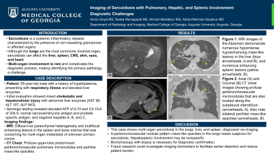
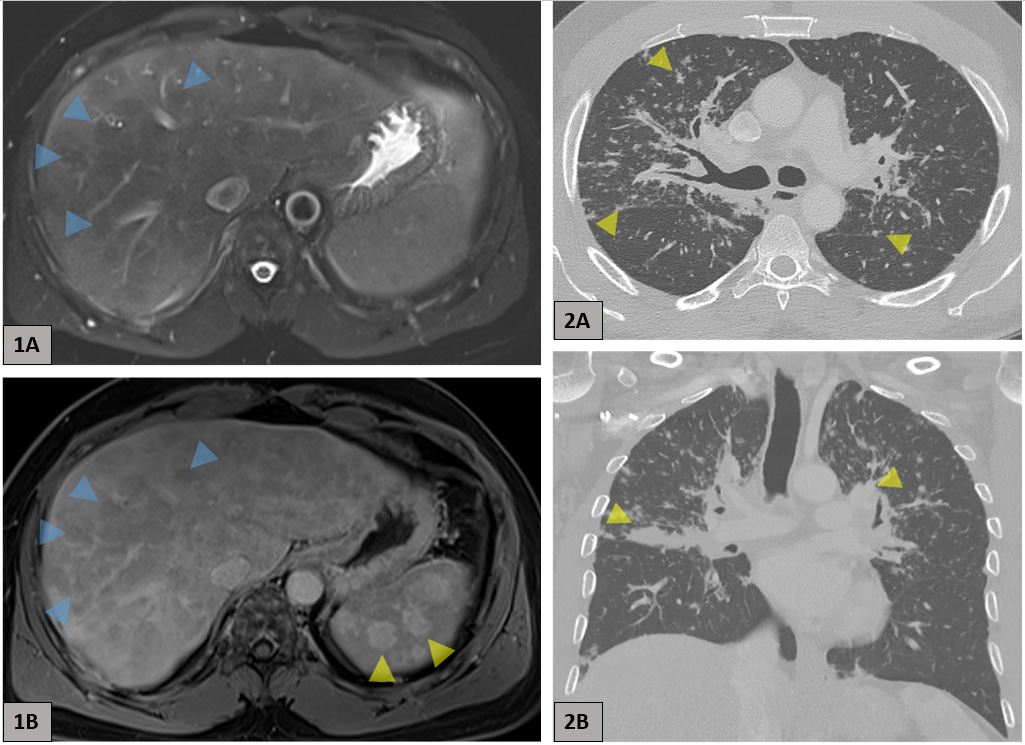
Case Description/Methods: A 55-year-old male with hyperlipidemia was evaluated for respiratory illness with elevated alkaline phosphatase (ALP) at an outside hospital. Further testing revealed elevated aspartate aminotransferase (AST) of 99, alanine aminotransferase (ALT) of 107, and ALP of 843, suggestive of mixed cholestatic and hepatocellular injury. He was referred to gastroenterology for evaluation of abnormal liver enzymes. At the time of presentation, the patient denied abdominal pain and jaundice. Serology showed hepatitis A/B immunity, negative hepatitis C, and normal ceruloplasmin, anti-mitochondrial and anti-smooth muscle antibodies, along with normal blood count and electrolytes. MRI showed diffuse liver parenchymal heterogeneity and multifocal enhancing lesions in the spleen and bone marrow that was concerning for multi-organ metastasis of unknown primary cancer (Figure 1). Oncology recommended tumor markers evaluation and a chest CT scan. Serology revealed elevated alpha-fetoprotein of 9.74 and CA 19-9 of 204.9 with normal carcinoembryonic antigen and prostate specific antigen. CT chest showed profuse upper-lobe predominant peribronchovascular pulmonary micronodules and perihilar mass-like opacities that is typical for pulmonary sarcoidosis (Figure 2). Bronchoscopy and biopsy is pending.
Discussion: This case highlights multi-organ sarcoidosis affecting the lungs, liver, and spleen, diagnosed through imaging. Although manifestations of liver and spleen involvement are nonspecific, their presence alongside a peribronchovascular nodular pattern and mass-like opacities in the lungs raises suspicion for sarcoidosis. Histology remains essential for definitive diagnosis, while future research could investigate imaging biomarkers to facilitate earlier detection and reduce patient burden.

Disclosures:
Arnav Goyal, BS1, Sweta Munagapati, BS1, Ahmad Alkaddour, MD1, Abdul-Rahman Abualruz, MD2. P3002 - Imaging of Sarcoidosis With Pulmonary, Hepatic, and Splenic Involvement: Diagnostic Challenges, ACG 2024 Annual Scientific Meeting Abstracts. Philadelphia, PA: American College of Gastroenterology.
1Medical College of Georgia at Augusta University, Augusta, GA; 2Medical College of Georgia, Augusta University, Augusta, GA
Introduction: Sarcoidosis is a systemic inflammatory disease characterized by non-caseating granulomas in various organs. While lungs are typically involved, sarcoidosis can affect the liver, spleen, CNS, skin, eyes, and heart. Multi-organ sarcoidosis is rare and presents diagnostic difficulties.
Case Description/Methods: A 55-year-old male with hyperlipidemia was evaluated for respiratory illness with elevated alkaline phosphatase (ALP) at an outside hospital. Further testing revealed elevated aspartate aminotransferase (AST) of 99, alanine aminotransferase (ALT) of 107, and ALP of 843, suggestive of mixed cholestatic and hepatocellular injury. He was referred to gastroenterology for evaluation of abnormal liver enzymes. At the time of presentation, the patient denied abdominal pain and jaundice. Serology showed hepatitis A/B immunity, negative hepatitis C, and normal ceruloplasmin, anti-mitochondrial and anti-smooth muscle antibodies, along with normal blood count and electrolytes. MRI showed diffuse liver parenchymal heterogeneity and multifocal enhancing lesions in the spleen and bone marrow that was concerning for multi-organ metastasis of unknown primary cancer (Figure 1). Oncology recommended tumor markers evaluation and a chest CT scan. Serology revealed elevated alpha-fetoprotein of 9.74 and CA 19-9 of 204.9 with normal carcinoembryonic antigen and prostate specific antigen. CT chest showed profuse upper-lobe predominant peribronchovascular pulmonary micronodules and perihilar mass-like opacities that is typical for pulmonary sarcoidosis (Figure 2). Bronchoscopy and biopsy is pending.
Discussion: This case highlights multi-organ sarcoidosis affecting the lungs, liver, and spleen, diagnosed through imaging. Although manifestations of liver and spleen involvement are nonspecific, their presence alongside a peribronchovascular nodular pattern and mass-like opacities in the lungs raises suspicion for sarcoidosis. Histology remains essential for definitive diagnosis, while future research could investigate imaging biomarkers to facilitate earlier detection and reduce patient burden.

Figure: Figure 1: MRI images of the Abdomen demonstrate numerous hypointense hypoenhancing mass-like lesions in the liver (blue arrowheads, A and B), and numerous enhancing splenic lesions (yellow arrowheads, B).
Figure 2: Axial (A) and Coronal (B) CT chest images showing profuse peribronchovascular micronodules that are also located along the subpleural interstitium (arrowheads, A). Also note bilateral perihilar mass-like opacities (arrowheads, B).
Figure 2: Axial (A) and Coronal (B) CT chest images showing profuse peribronchovascular micronodules that are also located along the subpleural interstitium (arrowheads, A). Also note bilateral perihilar mass-like opacities (arrowheads, B).
Disclosures:
Arnav Goyal indicated no relevant financial relationships.
Sweta Munagapati indicated no relevant financial relationships.
Ahmad Alkaddour indicated no relevant financial relationships.
Abdul-Rahman Abualruz indicated no relevant financial relationships.
Arnav Goyal, BS1, Sweta Munagapati, BS1, Ahmad Alkaddour, MD1, Abdul-Rahman Abualruz, MD2. P3002 - Imaging of Sarcoidosis With Pulmonary, Hepatic, and Splenic Involvement: Diagnostic Challenges, ACG 2024 Annual Scientific Meeting Abstracts. Philadelphia, PA: American College of Gastroenterology.
