Monday Poster Session
Category: Liver
P3042 - A Case of Non-Occlusive Mesenteric Ischemia Following the Rupture of a Hepatocellular Carcinoma
Monday, October 28, 2024
10:30 AM - 4:00 PM ET
Location: Exhibit Hall E

Has Audio
- IR
Ivana Radosavljevic, MD
University of South Florida Morsani College of Medicine
Tampa, FL
Presenting Author(s)
Ivana Radosavljevic, MD1, Takao Miwa, MD, PhD2, Masafumi Kawade, MD2, Shinji Unome, MD2, Kenji Imai, MD2, Koji Takai, MD2, Masahito Shimizu, MD2
1University of South Florida Morsani College of Medicine, Tampa, FL; 2Gifu University, Gifu, Gifu, Japan
Introduction: Hepatocellular carcinoma (HCC) rupture rates are increasing, with Asian regions reaching an incidence of 15%. Although rupture of HCC can cause various complications, there is little literature available on patients who develop nonocclusive mesenteric ischemia (NOMI) following HCC rupture. Herein, we report a case of HCC rupture with subsequent NOMI that was successfully managed.
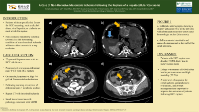
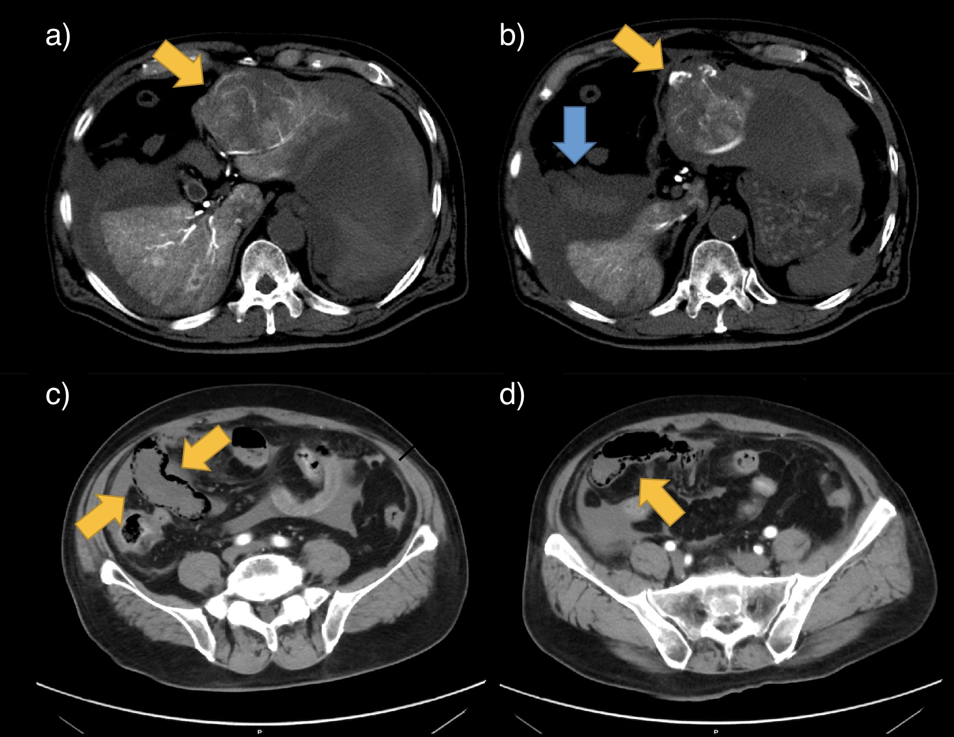
Case Description/Methods: A 77-year-old otherwise healthy man with no history of smoking or alcohol abuse presented to an emergency department due to progressively worsening abdominal pain. After a computed topography (CT) scan showed a liver tumor with lateral abdominal ascites, he was transferred to our hospital for a suspected HCC rupture. On arrival, his vital signs showed hemodynamic instability and his physical exam revealed conjunctival pallor, spontaneous abdominal pain, and tenderness to palpation throughout his abdomen. Significant laboratory findings included a low hemoglobin of 7.6 g/dL and elevated levels of prothrombin induced by vitamin K absence or antagonist-II, an HCC tumor marker, of 3612 mAU/ml. The patient underwent a hepatic artery embolization procedure. The arterial phase of the CT hepatic arteriography a slightly enhanced 77 mm hepatocellular carcinoma in in the left lobe (Figure 1a) with extravasation (Figure 1b). Therefore, the left ventro-lateral branch was embolized using gelatin sponge. The next day, he reported abdominal pain and laboratory data showed a marked lactic acidosis. A CT scan with contrast showed pneumatosis intestinalis and reduced enhancement in the small intestine (Figure 1c, 1d). A small intestine resection was performed and histology confirmed hemorrhage, congestion, necrosis, and diffuse inflammatory cell infiltration within the intestinal wall without abnormal findings in the mesenteric vessels, which was consistent with the clinical diagnosis of NOMI. The patient gradually recovered and was discharged after rehabilitation.
Discussion: This is the first report of HCC rupture leading to NOMI. Given the high mortality of these conditions, this case has significant implications for the diagnosis and management of NOMI in this population. A high level of suspicion for complications, comprehensive evaluations, and prompt intervention can lead to improved outcomes in patients with HCC rupture.

Disclosures:
Ivana Radosavljevic, MD1, Takao Miwa, MD, PhD2, Masafumi Kawade, MD2, Shinji Unome, MD2, Kenji Imai, MD2, Koji Takai, MD2, Masahito Shimizu, MD2. P3042 - A Case of Non-Occlusive Mesenteric Ischemia Following the Rupture of a Hepatocellular Carcinoma, ACG 2024 Annual Scientific Meeting Abstracts. Philadelphia, PA: American College of Gastroenterology.
1University of South Florida Morsani College of Medicine, Tampa, FL; 2Gifu University, Gifu, Gifu, Japan
Introduction: Hepatocellular carcinoma (HCC) rupture rates are increasing, with Asian regions reaching an incidence of 15%. Although rupture of HCC can cause various complications, there is little literature available on patients who develop nonocclusive mesenteric ischemia (NOMI) following HCC rupture. Herein, we report a case of HCC rupture with subsequent NOMI that was successfully managed.
Case Description/Methods: A 77-year-old otherwise healthy man with no history of smoking or alcohol abuse presented to an emergency department due to progressively worsening abdominal pain. After a computed topography (CT) scan showed a liver tumor with lateral abdominal ascites, he was transferred to our hospital for a suspected HCC rupture. On arrival, his vital signs showed hemodynamic instability and his physical exam revealed conjunctival pallor, spontaneous abdominal pain, and tenderness to palpation throughout his abdomen. Significant laboratory findings included a low hemoglobin of 7.6 g/dL and elevated levels of prothrombin induced by vitamin K absence or antagonist-II, an HCC tumor marker, of 3612 mAU/ml. The patient underwent a hepatic artery embolization procedure. The arterial phase of the CT hepatic arteriography a slightly enhanced 77 mm hepatocellular carcinoma in in the left lobe (Figure 1a) with extravasation (Figure 1b). Therefore, the left ventro-lateral branch was embolized using gelatin sponge. The next day, he reported abdominal pain and laboratory data showed a marked lactic acidosis. A CT scan with contrast showed pneumatosis intestinalis and reduced enhancement in the small intestine (Figure 1c, 1d). A small intestine resection was performed and histology confirmed hemorrhage, congestion, necrosis, and diffuse inflammatory cell infiltration within the intestinal wall without abnormal findings in the mesenteric vessels, which was consistent with the clinical diagnosis of NOMI. The patient gradually recovered and was discharged after rehabilitation.
Discussion: This is the first report of HCC rupture leading to NOMI. Given the high mortality of these conditions, this case has significant implications for the diagnosis and management of NOMI in this population. A high level of suspicion for complications, comprehensive evaluations, and prompt intervention can lead to improved outcomes in patients with HCC rupture.

Figure: Hepatic arteriography (a, b) showing a slightly enhanced hepatocellular carcinoma (yellow arrow) in in the left lobe with with extravasation (yellow arrow) and hemorrhagic ascites (blue arrow). An abdominal contrast-enhanced computed tomography coronal section (c, d) revealed pneumatosis intestinalis and reduced enhancement in the wall of the small intestine.
Disclosures:
Ivana Radosavljevic indicated no relevant financial relationships.
Takao Miwa indicated no relevant financial relationships.
Masafumi Kawade indicated no relevant financial relationships.
Shinji Unome indicated no relevant financial relationships.
Kenji Imai indicated no relevant financial relationships.
Koji Takai indicated no relevant financial relationships.
Masahito Shimizu indicated no relevant financial relationships.
Ivana Radosavljevic, MD1, Takao Miwa, MD, PhD2, Masafumi Kawade, MD2, Shinji Unome, MD2, Kenji Imai, MD2, Koji Takai, MD2, Masahito Shimizu, MD2. P3042 - A Case of Non-Occlusive Mesenteric Ischemia Following the Rupture of a Hepatocellular Carcinoma, ACG 2024 Annual Scientific Meeting Abstracts. Philadelphia, PA: American College of Gastroenterology.

