Monday Poster Session
Category: Liver
P3049 - Beyond the Routine: Unusual Abdominal Pseudocyst Following Peritoneal Dialysis
Monday, October 28, 2024
10:30 AM - 4:00 PM ET
Location: Exhibit Hall E

Has Audio
.jpg)
Natasha Mederos, MD
University of Miami Miller School of Medicine at Holy Cross Hospital, FL
Presenting Author(s)
Natasha Mederos, MD, John Coleman, MD, Mary Parianos, MD
University of Miami Miller School of Medicine at Holy Cross Hospital, Fort Lauderdale, FL
Introduction: Peritoneal dialysis (PD) offers numerous advantages, yet complications such as leakage, peritonitis, and abdominal wall weakness can emerge. Rarely reported are cases of abdominal and peritoneal pseudocyst formation as a PD complication, typically in the inflammatory context. Non-pancreatic intra-abdominal pseudocysts, with an incidence of approximately 0.0001% to 0.000004% of hospital admissions, usually link to major intra-abdominal surgeries, trauma, ventriculoperitoneal (VP) shunts, or intra-peritoneal catheter infections.
Case Description/Methods: A 55-year-old male with a history of hypertension and end stage renal disease on hemodialysis presented to the emergency department with progressive abdominal discomfort and distention accompanied by reflux and back pain that started months prior to admission. He denied associated symptoms, such as nausea, vomiting, constipation, diarrhea, weight changes, fevers, or chills. Patient had been switched from continuous peritoneal dialysis to hemodialysis six months prior due to poor output and bloody drainage, yet retained the PD catheter in the hope of returning to peritoneal dialysis.
On arrival, patient was found to have a distended, hard, non-tender abdomen with decreased bowel sounds on physical examination. A computed tomography (CT) abdomen with contrast revelead a large thick-walled fluid collection versus mass lesion measuring 27 x 26 x 23 cm. CT-guided percutaneous drainage was performed, yielding 6 L of old bloody material. Follow-up imaging with CT abdomen scan indicated a residual smaller cystic lesion. Surgical intervention for removal of the peritoneal dialysis catheter, excisional debridement of the abdominal wound, and laparoscopic drainage of the cystic mass were conducted. The cyst was firmly adherent to the abdominal wall, posed challenges for complete removal, leading to the decision for complete drainage rather than surgical excision. The cystic mass was non-malignant, and the patient was safely discharged home after post-operative recovery.
Discussion: Surgical intervention for peritoneal pseudocyst is considered curative; however, comorbidities heighten surgical risks, and infection poses a risk for pseudocyst formation. In cases where the pseudocyst is inoperable, IR-guided drainage serves as an alternative, despite increasing the likelihood of recurrence. These insights underscore the need for close follow-up in managing abdominal cystic lesions.

Disclosures:
Natasha Mederos, MD, John Coleman, MD, Mary Parianos, MD. P3049 - Beyond the Routine: Unusual Abdominal Pseudocyst Following Peritoneal Dialysis, ACG 2024 Annual Scientific Meeting Abstracts. Philadelphia, PA: American College of Gastroenterology.
University of Miami Miller School of Medicine at Holy Cross Hospital, Fort Lauderdale, FL
Introduction: Peritoneal dialysis (PD) offers numerous advantages, yet complications such as leakage, peritonitis, and abdominal wall weakness can emerge. Rarely reported are cases of abdominal and peritoneal pseudocyst formation as a PD complication, typically in the inflammatory context. Non-pancreatic intra-abdominal pseudocysts, with an incidence of approximately 0.0001% to 0.000004% of hospital admissions, usually link to major intra-abdominal surgeries, trauma, ventriculoperitoneal (VP) shunts, or intra-peritoneal catheter infections.
Case Description/Methods: A 55-year-old male with a history of hypertension and end stage renal disease on hemodialysis presented to the emergency department with progressive abdominal discomfort and distention accompanied by reflux and back pain that started months prior to admission. He denied associated symptoms, such as nausea, vomiting, constipation, diarrhea, weight changes, fevers, or chills. Patient had been switched from continuous peritoneal dialysis to hemodialysis six months prior due to poor output and bloody drainage, yet retained the PD catheter in the hope of returning to peritoneal dialysis.
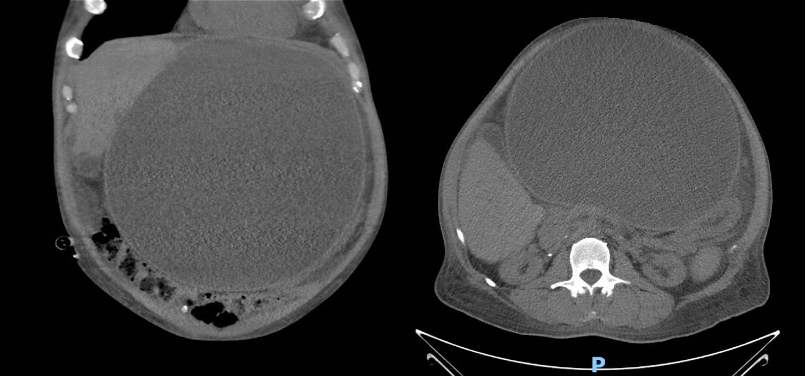
On arrival, patient was found to have a distended, hard, non-tender abdomen with decreased bowel sounds on physical examination. A computed tomography (CT) abdomen with contrast revelead a large thick-walled fluid collection versus mass lesion measuring 27 x 26 x 23 cm. CT-guided percutaneous drainage was performed, yielding 6 L of old bloody material. Follow-up imaging with CT abdomen scan indicated a residual smaller cystic lesion. Surgical intervention for removal of the peritoneal dialysis catheter, excisional debridement of the abdominal wound, and laparoscopic drainage of the cystic mass were conducted. The cyst was firmly adherent to the abdominal wall, posed challenges for complete removal, leading to the decision for complete drainage rather than surgical excision. The cystic mass was non-malignant, and the patient was safely discharged home after post-operative recovery.
Discussion: Surgical intervention for peritoneal pseudocyst is considered curative; however, comorbidities heighten surgical risks, and infection poses a risk for pseudocyst formation. In cases where the pseudocyst is inoperable, IR-guided drainage serves as an alternative, despite increasing the likelihood of recurrence. These insights underscore the need for close follow-up in managing abdominal cystic lesions.

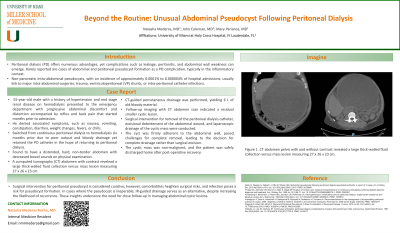
Figure: Figure 1. CT abdomen pelvis with and without contrast revealed a large thick-walled fluid collection versus mass lesion measuring 27 x 26 x 23 cm.
Disclosures:
Natasha Mederos indicated no relevant financial relationships.
John Coleman indicated no relevant financial relationships.
Mary Parianos indicated no relevant financial relationships.
Natasha Mederos, MD, John Coleman, MD, Mary Parianos, MD. P3049 - Beyond the Routine: Unusual Abdominal Pseudocyst Following Peritoneal Dialysis, ACG 2024 Annual Scientific Meeting Abstracts. Philadelphia, PA: American College of Gastroenterology.
