Monday Poster Session
Category: Liver
P3051 - Silent Hemorrhage: Spontaneous Muscle Hematoma in Liver Cirrhosis Unveiled
Monday, October 28, 2024
10:30 AM - 4:00 PM ET
Location: Exhibit Hall E

Has Audio

Warsha Mal Korani, MBBS
Conemaugh Memorial Medical Center
JOHNSTOWN, PA
Presenting Author(s)
Warsha Mal Korani, MBBS1, Pan Zonghao, MD2, Muhammad Adnan Zaman, MBBS1, Hussain Khandakar, MD1
1Conemaugh Memorial Medical Center, Johnstown, PA; 2Temple University/Conemaugh Memorial Medical Center Internal Medicine, Johnstown, PA
Introduction: In 2016, cirrhosis caused 34,174 deaths in the US, primarily due to variceal hemorrhage, infections, cancer, and renal failure. Among these complications, spontaneous rectus sheath hematomas are infrequently reported. This report details a case of a cirrhotic patient who succumbed to a spontaneous RSH.
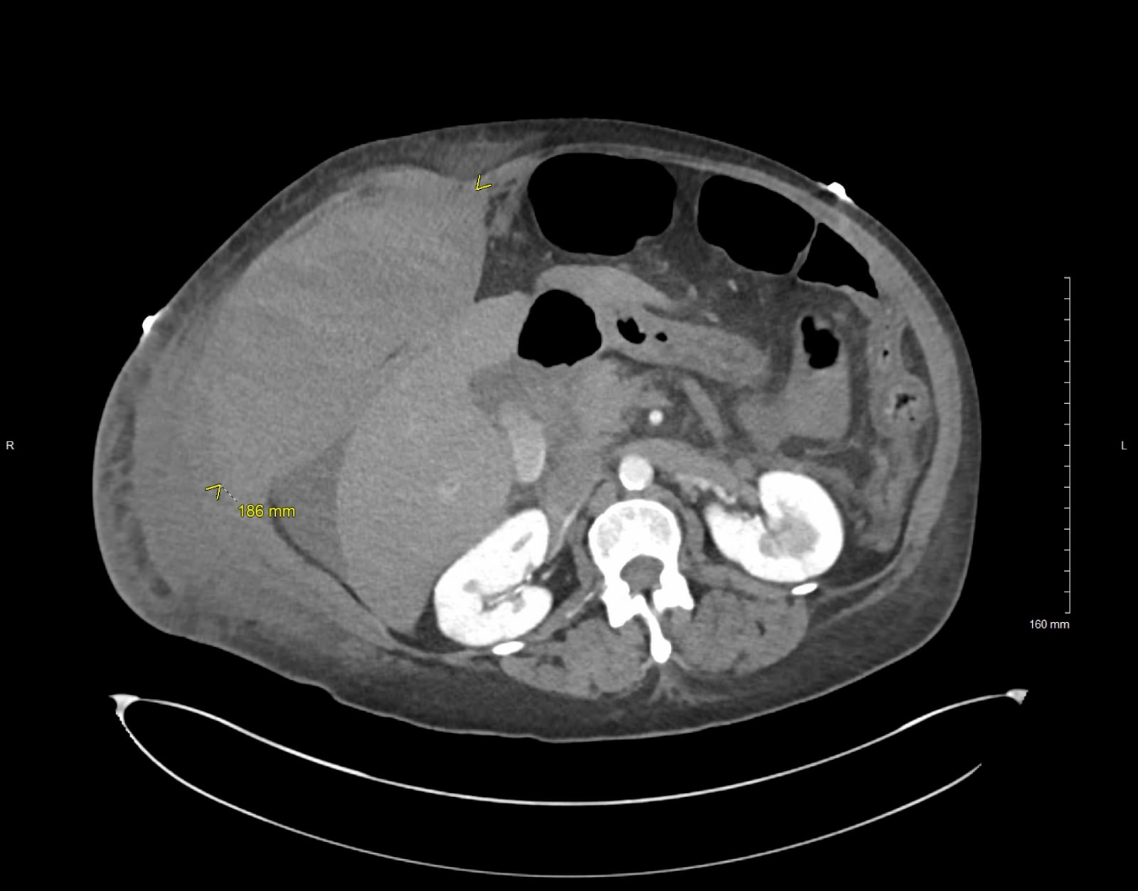
Case Description/Methods: A 60-year-old woman with alcohol use disorder, untreated hepatitis C, and newly diagnosed liver cirrhosis presented with severe right upper quadrant pain. Initial labs showed WBC 13 × 10^9/L, Hgb 10 g/dL, sodium 127 mmol/L, creatinine 1.2 mg/dL, total bilirubin 7.0 mg/dL, direct bilirubin 3.7 mg/dL, AST 75 U/L, ALT 22 U/L, albumin 2.9 g/dL, INR 2.1, PT 22 seconds, and lactate 3.0 mmol/L. CT scan revealed hepatic cirrhosis with varices and moderate ascites. She was admitted for decompensated liver cirrhosis. On the third day, she developed worsening abdominal pain and hematemesis, with hemoglobin dropping from 10.0 to 6.2 g/dL, necessitating urgent blood transfusion and octreotide infusion due to suspected variceal bleeding. Emergent imaging showed an acute rectus sheath hematoma with active bleeding from the right anterior abdominal wall. Interventional radiology performed embolization of the right circumflex artery branch, initially controlling the bleed. Despite these measures, she experienced persistent bleeding, leading to DIC, AKI, and recurrent bleeding from the right inferior epigastric artery, right deep circumflex iliac artery, and distal right internal thoracic/superior epigastric artery, all of which were subsequently embolized. Despite aggressive treatment, she developed severe metabolic acidosis, had five episodes of PEA, and ultimately succumbed to her condition.
Discussion: Spontaneous rectus sheath hematomas, though rare, pose critical risks. Factors like anticoagulant use, advanced kidney disease, hypertension, age, recent abdominal procedures increase susceptibility. Cirrhosis exacerbates bleeding risk due to liver dysfunction, thrombocytopenia, and splenomegaly, yet RSHs in cirrhotic patients are seldom reported. Symptoms include abdominal pain, palpable mass, expanding girth,ecchymosis, and hypotension. Initial treatment entails correction of coagulopathy. Transarterial embolization, generally safe and effective, may require multiple attempts.Refractory cases might benefit from a transjugular intrahepatic portosystemic shunt. Prompt recognition and aggressive management are crucial to improve outcomes in this vulnerable population

Disclosures:
Warsha Mal Korani, MBBS1, Pan Zonghao, MD2, Muhammad Adnan Zaman, MBBS1, Hussain Khandakar, MD1. P3051 - Silent Hemorrhage: Spontaneous Muscle Hematoma in Liver Cirrhosis Unveiled, ACG 2024 Annual Scientific Meeting Abstracts. Philadelphia, PA: American College of Gastroenterology.
1Conemaugh Memorial Medical Center, Johnstown, PA; 2Temple University/Conemaugh Memorial Medical Center Internal Medicine, Johnstown, PA
Introduction: In 2016, cirrhosis caused 34,174 deaths in the US, primarily due to variceal hemorrhage, infections, cancer, and renal failure. Among these complications, spontaneous rectus sheath hematomas are infrequently reported. This report details a case of a cirrhotic patient who succumbed to a spontaneous RSH.
Case Description/Methods: A 60-year-old woman with alcohol use disorder, untreated hepatitis C, and newly diagnosed liver cirrhosis presented with severe right upper quadrant pain. Initial labs showed WBC 13 × 10^9/L, Hgb 10 g/dL, sodium 127 mmol/L, creatinine 1.2 mg/dL, total bilirubin 7.0 mg/dL, direct bilirubin 3.7 mg/dL, AST 75 U/L, ALT 22 U/L, albumin 2.9 g/dL, INR 2.1, PT 22 seconds, and lactate 3.0 mmol/L. CT scan revealed hepatic cirrhosis with varices and moderate ascites. She was admitted for decompensated liver cirrhosis. On the third day, she developed worsening abdominal pain and hematemesis, with hemoglobin dropping from 10.0 to 6.2 g/dL, necessitating urgent blood transfusion and octreotide infusion due to suspected variceal bleeding. Emergent imaging showed an acute rectus sheath hematoma with active bleeding from the right anterior abdominal wall. Interventional radiology performed embolization of the right circumflex artery branch, initially controlling the bleed. Despite these measures, she experienced persistent bleeding, leading to DIC, AKI, and recurrent bleeding from the right inferior epigastric artery, right deep circumflex iliac artery, and distal right internal thoracic/superior epigastric artery, all of which were subsequently embolized. Despite aggressive treatment, she developed severe metabolic acidosis, had five episodes of PEA, and ultimately succumbed to her condition.
Discussion: Spontaneous rectus sheath hematomas, though rare, pose critical risks. Factors like anticoagulant use, advanced kidney disease, hypertension, age, recent abdominal procedures increase susceptibility. Cirrhosis exacerbates bleeding risk due to liver dysfunction, thrombocytopenia, and splenomegaly, yet RSHs in cirrhotic patients are seldom reported. Symptoms include abdominal pain, palpable mass, expanding girth,ecchymosis, and hypotension. Initial treatment entails correction of coagulopathy. Transarterial embolization, generally safe and effective, may require multiple attempts.Refractory cases might benefit from a transjugular intrahepatic portosystemic shunt. Prompt recognition and aggressive management are crucial to improve outcomes in this vulnerable population

Figure: Right abdominal wall Hematoma
Disclosures:
Warsha Mal Korani indicated no relevant financial relationships.
Pan Zonghao indicated no relevant financial relationships.
Muhammad Adnan Zaman indicated no relevant financial relationships.
Hussain Khandakar indicated no relevant financial relationships.
Warsha Mal Korani, MBBS1, Pan Zonghao, MD2, Muhammad Adnan Zaman, MBBS1, Hussain Khandakar, MD1. P3051 - Silent Hemorrhage: Spontaneous Muscle Hematoma in Liver Cirrhosis Unveiled, ACG 2024 Annual Scientific Meeting Abstracts. Philadelphia, PA: American College of Gastroenterology.
