Monday Poster Session
Category: Liver
P3071 - A Rare Presentation of a Gastrointestinal Neuroendocrine Tumor with Ascites and Negative Cytology: A Case Report
Monday, October 28, 2024
10:30 AM - 4:00 PM ET
Location: Exhibit Hall E

Has Audio
- WH
We'am Hussain, MD
Wright State University Boonshoft School of Medicine
Dayton, OH
Presenting Author(s)
We'am Hussain, MD1, Omair Khan, MS2, Shashank Mittal, MD1, Asif Jan, MD3, Kaylee Larsen, MS1, Maaz Arif, MD1, Sangeeta Agrawal, MD3
1Wright State University Boonshoft School of Medicine, Dayton, OH; 2Kansas City University, Dayton, OH; 3Wright State University, Dayton, OH
Introduction: Neuroendocrine tumors are a rare malignancy that make up 0.5% of all malignancy diagnoses with < 200,000 cases in the United States. They can present in a wide variety of anatomic locations and produce a diversity of clinical syndromes; thus diagnosis often requires tissue histology.
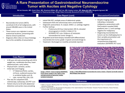
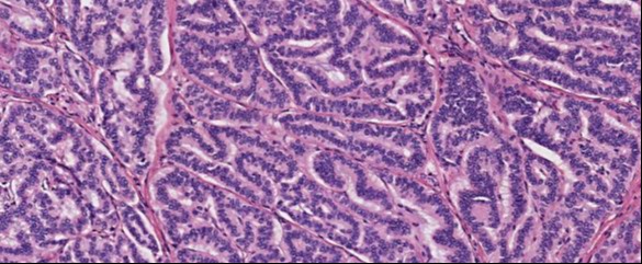
Case Description/Methods: A 69-year-old man presented with 40-pound unintentional weight loss, non-bloody diarrhea, lower abdominal pain, and early satiety. Cachexia was noted on the exam. CT of the abdomen showed ascites without cirrhosis and scattered sclerotic osseous foci within multiple vertebral bodies and a rib, which was overall concerning for malignancy. Outpatient paracentesis by IR yielded 3.7 L fluid output, for which cell studies were negative for malignancy. Prostate specific antigen (PSA), lactate dehydrogenase (LDH), and carcinoembryonic antigen (CEA) labs were normal. A plan was then formulated for further workup including EGD, colonoscopy, cystoscopy, and PET scan. However, the patient soon returned to the ED with diffuse abdominal pain and distention. CT of the abdomen demonstrated re-accumulation of ascites. Labs were notable for hypoalbuminemia without gamma gap elevation, and he was admitted for further workup. Initial fludeoxyglucose-18 (FDG) PET scan showed multiple areas of uptake in the abdomen. Despite this, EGD and colonoscopy were unremarkable, and CA 19-9 and PSA labs were normal. He underwent diagnostic paracentesis by GI, yielding 5 L fluid output. Serum and fluid studies were notable for SAAG of 1.2, hypoproteinemia (serum total protein of 4.8), and cytology negative for malignancy (Table 1). Further studies were notable for proteinuria (24-hour urine total protein 205.5), elevated chromogranin A level (1419), and elevated 5-HIAA level (17.5). A DOTATATE PET scan was performed demonstrating evidence of metastatic neuroendocrine tumor. Evaluations by IR and surgery demonstrated no suitable soft tissue biopsy location, so bone scan was performed. Consequently, IR-guided sacral bone biopsy was performed, and pathology showed a well-differentiated neuroendocrine tumor, grade 1 (typical carcinoid). The patient was then started on octreotide for metastatic neuroendocrine tumor. The patient was discharged on monthly octreotide injections with a plan to repeat DOTATATE PET scan in a few months.
Discussion: Despite evidence of metastatic malignancy on CT, PET, and bone scans, numerous paracenteses with fluid analyses yielded SAAG gradients over 1.1 and no evidence of malignancy on cytology.

Note: The table for this abstract can be viewed in the ePoster Gallery section of the ACG 2024 ePoster Site or in The American Journal of Gastroenterology's abstract supplement issue, both of which will be available starting October 27, 2024.
Disclosures:
We'am Hussain, MD1, Omair Khan, MS2, Shashank Mittal, MD1, Asif Jan, MD3, Kaylee Larsen, MS1, Maaz Arif, MD1, Sangeeta Agrawal, MD3. P3071 - A Rare Presentation of a Gastrointestinal Neuroendocrine Tumor with Ascites and Negative Cytology: A Case Report, ACG 2024 Annual Scientific Meeting Abstracts. Philadelphia, PA: American College of Gastroenterology.
1Wright State University Boonshoft School of Medicine, Dayton, OH; 2Kansas City University, Dayton, OH; 3Wright State University, Dayton, OH
Introduction: Neuroendocrine tumors are a rare malignancy that make up 0.5% of all malignancy diagnoses with < 200,000 cases in the United States. They can present in a wide variety of anatomic locations and produce a diversity of clinical syndromes; thus diagnosis often requires tissue histology.
Case Description/Methods: A 69-year-old man presented with 40-pound unintentional weight loss, non-bloody diarrhea, lower abdominal pain, and early satiety. Cachexia was noted on the exam. CT of the abdomen showed ascites without cirrhosis and scattered sclerotic osseous foci within multiple vertebral bodies and a rib, which was overall concerning for malignancy. Outpatient paracentesis by IR yielded 3.7 L fluid output, for which cell studies were negative for malignancy. Prostate specific antigen (PSA), lactate dehydrogenase (LDH), and carcinoembryonic antigen (CEA) labs were normal. A plan was then formulated for further workup including EGD, colonoscopy, cystoscopy, and PET scan. However, the patient soon returned to the ED with diffuse abdominal pain and distention. CT of the abdomen demonstrated re-accumulation of ascites. Labs were notable for hypoalbuminemia without gamma gap elevation, and he was admitted for further workup. Initial fludeoxyglucose-18 (FDG) PET scan showed multiple areas of uptake in the abdomen. Despite this, EGD and colonoscopy were unremarkable, and CA 19-9 and PSA labs were normal. He underwent diagnostic paracentesis by GI, yielding 5 L fluid output. Serum and fluid studies were notable for SAAG of 1.2, hypoproteinemia (serum total protein of 4.8), and cytology negative for malignancy (Table 1). Further studies were notable for proteinuria (24-hour urine total protein 205.5), elevated chromogranin A level (1419), and elevated 5-HIAA level (17.5). A DOTATATE PET scan was performed demonstrating evidence of metastatic neuroendocrine tumor. Evaluations by IR and surgery demonstrated no suitable soft tissue biopsy location, so bone scan was performed. Consequently, IR-guided sacral bone biopsy was performed, and pathology showed a well-differentiated neuroendocrine tumor, grade 1 (typical carcinoid). The patient was then started on octreotide for metastatic neuroendocrine tumor. The patient was discharged on monthly octreotide injections with a plan to repeat DOTATATE PET scan in a few months.
Discussion: Despite evidence of metastatic malignancy on CT, PET, and bone scans, numerous paracenteses with fluid analyses yielded SAAG gradients over 1.1 and no evidence of malignancy on cytology.

Figure: Image 1: Well differentiated neuroendocrine tumor, grade 1 (Typical Carcinoid) noted on Bone Biopsy from Sacrum
Note: The table for this abstract can be viewed in the ePoster Gallery section of the ACG 2024 ePoster Site or in The American Journal of Gastroenterology's abstract supplement issue, both of which will be available starting October 27, 2024.
Disclosures:
We'am Hussain indicated no relevant financial relationships.
Omair Khan indicated no relevant financial relationships.
Shashank Mittal indicated no relevant financial relationships.
Asif Jan indicated no relevant financial relationships.
Kaylee Larsen indicated no relevant financial relationships.
Maaz Arif indicated no relevant financial relationships.
Sangeeta Agrawal indicated no relevant financial relationships.
We'am Hussain, MD1, Omair Khan, MS2, Shashank Mittal, MD1, Asif Jan, MD3, Kaylee Larsen, MS1, Maaz Arif, MD1, Sangeeta Agrawal, MD3. P3071 - A Rare Presentation of a Gastrointestinal Neuroendocrine Tumor with Ascites and Negative Cytology: A Case Report, ACG 2024 Annual Scientific Meeting Abstracts. Philadelphia, PA: American College of Gastroenterology.
