Monday Poster Session
Category: Liver
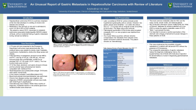
P3073 - An Unusual Report of Gastric Metastasis in Hepatocellular Carcinoma with Review of Literature
Monday, October 28, 2024
10:30 AM - 4:00 PM ET
Location: Exhibit Hall E

Has Audio
- NA
Narges Ashrafinia, MD
College of Medicine, University of Saskatchewan
Edmonton, AB, Canada
Presenting Author(s)
Narges Ashrafinia, MD1, Mina Niazi, MD2
1College of Medicine, University of Saskatchewan, Saskatoon, SK, Canada; 2University of Saskatchewan, Saskatoon, SK, Canada
Introduction: Hepatocellular carcinoma (HCC) is a primary neoplasm of the liver that develops in a setting of chronic liver disease and cirrhosis. Metastasis in the stomach is extremely rare. We present a case of HCC, secondary to metabolic dysfunction-associated steatohepatitis (MASH)-related cirrhosis, with an incidental finding of gastric metastasis during variceal surveillance.
Case Description/Methods: A 72-year-old man presented to the Emergency Department with acute onset abdominal pain. His medical history was notable for stroke, type-2 diabetes, hypertension, dyslipidemia, obstructive sleep apnea and obesity. Initial laboratory investigations showed normal levels of leukocytes and platelets and a hemoglobin level of 104 g/L (126-180 g/L). His liver enzyme panel was unremarkable, except for an elevated γ-glutamyl transferase level of 1.82 ukat/L (0.07-1 ukat/L). The liver function tests were normal. Computed tomography (CT) of the chest, abdomen, and pelvis illustrated cirrhosis and two arterial hyper-enhancing liver lesions compatible with multifocal HCC. Alpha-fetoprotein and carbohydrate antigen 19-9 levels were within normal limits. A liver biopsy revealed a well-differentiated HCC. Subsequently, trans-arterial chemoembolization was performed. Chronic liver disease workup was negative, and diagnosis of MASH compensated cirrhosis was made, with Child-Pugh A classification. Chemotherapy was initiated. Within three months of diagnosis, metastatic lesions in the adrenal gland and vertebral bodies were observed. Surveillance esophagogastroduodenoscopy (EGD) for varices showed portal hypertensive gastropathy with no varices. However, two gastric polyps were identified: 1 cm within the antrum and a pedunculated 2 × 3 cm polyp with an irregular border and pitting at the fundus. The larger polyp was removed. Pathological examination of the fundus polyp revealed metastatic HCC; no new symptom was reported from the patient. At a two-year follow-up period, cirrhosis remains compensated. His metastatic HCC extends to the splenic and pelvic osseous structures. The patient remains on chemotherapy.
Discussion: This case highlights the possibility of gastric metastasis in a patient with advanced HCC. The nonspecific presentation of gastric metastasis makes the diagnosis challenging. Hence, the supplementary role of EGD with meticulous inspection for abnormalities is crucial in diagnosing gastrointestinal metastasis.

Note: The table for this abstract can be viewed in the ePoster Gallery section of the ACG 2024 ePoster Site or in The American Journal of Gastroenterology's abstract supplement issue, both of which will be available starting October 27, 2024.
Disclosures:
Narges Ashrafinia, MD1, Mina Niazi, MD2. P3073 - An Unusual Report of Gastric Metastasis in Hepatocellular Carcinoma with Review of Literature, ACG 2024 Annual Scientific Meeting Abstracts. Philadelphia, PA: American College of Gastroenterology.
1College of Medicine, University of Saskatchewan, Saskatoon, SK, Canada; 2University of Saskatchewan, Saskatoon, SK, Canada
Introduction: Hepatocellular carcinoma (HCC) is a primary neoplasm of the liver that develops in a setting of chronic liver disease and cirrhosis. Metastasis in the stomach is extremely rare. We present a case of HCC, secondary to metabolic dysfunction-associated steatohepatitis (MASH)-related cirrhosis, with an incidental finding of gastric metastasis during variceal surveillance.
Case Description/Methods: A 72-year-old man presented to the Emergency Department with acute onset abdominal pain. His medical history was notable for stroke, type-2 diabetes, hypertension, dyslipidemia, obstructive sleep apnea and obesity. Initial laboratory investigations showed normal levels of leukocytes and platelets and a hemoglobin level of 104 g/L (126-180 g/L). His liver enzyme panel was unremarkable, except for an elevated γ-glutamyl transferase level of 1.82 ukat/L (0.07-1 ukat/L). The liver function tests were normal. Computed tomography (CT) of the chest, abdomen, and pelvis illustrated cirrhosis and two arterial hyper-enhancing liver lesions compatible with multifocal HCC. Alpha-fetoprotein and carbohydrate antigen 19-9 levels were within normal limits. A liver biopsy revealed a well-differentiated HCC. Subsequently, trans-arterial chemoembolization was performed. Chronic liver disease workup was negative, and diagnosis of MASH compensated cirrhosis was made, with Child-Pugh A classification. Chemotherapy was initiated. Within three months of diagnosis, metastatic lesions in the adrenal gland and vertebral bodies were observed. Surveillance esophagogastroduodenoscopy (EGD) for varices showed portal hypertensive gastropathy with no varices. However, two gastric polyps were identified: 1 cm within the antrum and a pedunculated 2 × 3 cm polyp with an irregular border and pitting at the fundus. The larger polyp was removed. Pathological examination of the fundus polyp revealed metastatic HCC; no new symptom was reported from the patient. At a two-year follow-up period, cirrhosis remains compensated. His metastatic HCC extends to the splenic and pelvic osseous structures. The patient remains on chemotherapy.
Discussion: This case highlights the possibility of gastric metastasis in a patient with advanced HCC. The nonspecific presentation of gastric metastasis makes the diagnosis challenging. Hence, the supplementary role of EGD with meticulous inspection for abnormalities is crucial in diagnosing gastrointestinal metastasis.

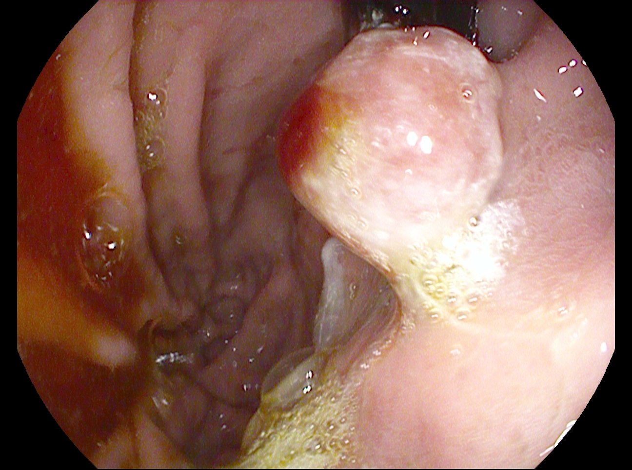
Figure: A pedunculated 2 x 3 cm polyp with central pitting and irregular border at the fundus.
Note: The table for this abstract can be viewed in the ePoster Gallery section of the ACG 2024 ePoster Site or in The American Journal of Gastroenterology's abstract supplement issue, both of which will be available starting October 27, 2024.
Disclosures:
Narges Ashrafinia indicated no relevant financial relationships.
Mina Niazi indicated no relevant financial relationships.
Narges Ashrafinia, MD1, Mina Niazi, MD2. P3073 - An Unusual Report of Gastric Metastasis in Hepatocellular Carcinoma with Review of Literature, ACG 2024 Annual Scientific Meeting Abstracts. Philadelphia, PA: American College of Gastroenterology.
