Monday Poster Session
Category: Small Intestine
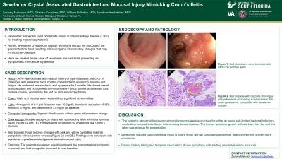
P3251 - Sevelamer Crystal Associated Gastrointestinal Mucosal Injury Mimicking Crohn’s Ileitis
Monday, October 28, 2024
10:30 AM - 4:00 PM ET
Location: Exhibit Hall E

Has Audio

Zachary Makovich, MD
University of South Florida Morsani College of Medicine
Tampa, FL
Presenting Author(s)
Zachary Makovich, MD1, Charles Cavalaris, MD1, Jonathan Keshishian, MD2, William Bulkeley, MD2
1University of South Florida Morsani College of Medicine, Tampa, FL; 2James A. Haley Veterans' Hospital, Tampa, FL
Introduction: Sevelamer is a phosphate binder used to treat hyperphosphatemia in patients with chronic kidney disease (CKD). In rare instances, sevelamer crystals can deposit within and disrupt the mucosa of the gastrointestinal tract resulting in bleeding and inflammatory changes that may mimic other diseases. We present a case of sevelamer induced ileitis presenting as symptomatic iron deficiency anemia.
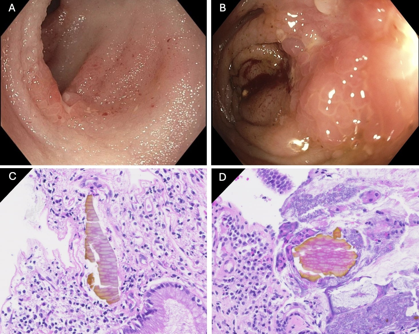
Case Description/Methods: A 76-year-old male with medical history of type II diabetes and CKD stage IV, notably on sevelamer for 3 months, presented to our hospital with worsening dyspnea and fatigue. He endorsed intermittent episodes of hematochezia and dyspepsia for 2 months. He denied use of anticoagulants or nonsteroidal anti-inflammatory drugs, unintentional weight loss, melena, nausea, or vomiting. He had no history of prior endoscopy. Vitals and physical exam were without significant abnormalities. Remarkable labs were a hemoglobin of 6.6 g/dl (baseline near 10.0 g/dl), transferrin saturation of 13%, ferritin of 27 ng/ml, and creatinine of 3.9 mg/dl (at baseline). Computed tomography showed sigmoid diverticulosis without gross inflammatory change. The patient was offered an esophagogastroduodenoscopy (EGD) and colonoscopy for work-up of severe iron deficiency anemia, however he declined the EGD, but did agree to colonoscopy. Colonoscopy performed without sedation revealed multiple serpiginous ulcers with surrounding ileitis within the terminal ileum (Figure 1A and 1B). Findings were concerning for underlying ileal Crohn’s disease. Ileal biopsies returned with focal reactive changes and pink and yellow crystalline material with a fish scale appearance, compatible with sevelamer crystals (Figure 1C and 1D). Findings were consistent with sevelamer crystal associated gastrointestinal mucosal injury. The patient’s sevelamer was discontinued, his gastrointestinal symptoms improved, and his hemoglobin improved to near baseline.
Discussion: The patient’s abnormalities seen during colonoscopy were suspicious for either an acute self-limited bacterial infection, medication induced enteritis, or inflammatory bowel disease. The former being incongruent with work up thus far, and the latter being atypical for presentation. Sevelamer induced ileitis is an underrecognized condition. This case underscores the importance of careful history taking and temporal association of new symptoms with starting new medications.

Disclosures:
Zachary Makovich, MD1, Charles Cavalaris, MD1, Jonathan Keshishian, MD2, William Bulkeley, MD2. P3251 - Sevelamer Crystal Associated Gastrointestinal Mucosal Injury Mimicking Crohn’s Ileitis, ACG 2024 Annual Scientific Meeting Abstracts. Philadelphia, PA: American College of Gastroenterology.
1University of South Florida Morsani College of Medicine, Tampa, FL; 2James A. Haley Veterans' Hospital, Tampa, FL
Introduction: Sevelamer is a phosphate binder used to treat hyperphosphatemia in patients with chronic kidney disease (CKD). In rare instances, sevelamer crystals can deposit within and disrupt the mucosa of the gastrointestinal tract resulting in bleeding and inflammatory changes that may mimic other diseases. We present a case of sevelamer induced ileitis presenting as symptomatic iron deficiency anemia.
Case Description/Methods: A 76-year-old male with medical history of type II diabetes and CKD stage IV, notably on sevelamer for 3 months, presented to our hospital with worsening dyspnea and fatigue. He endorsed intermittent episodes of hematochezia and dyspepsia for 2 months. He denied use of anticoagulants or nonsteroidal anti-inflammatory drugs, unintentional weight loss, melena, nausea, or vomiting. He had no history of prior endoscopy. Vitals and physical exam were without significant abnormalities. Remarkable labs were a hemoglobin of 6.6 g/dl (baseline near 10.0 g/dl), transferrin saturation of 13%, ferritin of 27 ng/ml, and creatinine of 3.9 mg/dl (at baseline). Computed tomography showed sigmoid diverticulosis without gross inflammatory change. The patient was offered an esophagogastroduodenoscopy (EGD) and colonoscopy for work-up of severe iron deficiency anemia, however he declined the EGD, but did agree to colonoscopy. Colonoscopy performed without sedation revealed multiple serpiginous ulcers with surrounding ileitis within the terminal ileum (Figure 1A and 1B). Findings were concerning for underlying ileal Crohn’s disease. Ileal biopsies returned with focal reactive changes and pink and yellow crystalline material with a fish scale appearance, compatible with sevelamer crystals (Figure 1C and 1D). Findings were consistent with sevelamer crystal associated gastrointestinal mucosal injury. The patient’s sevelamer was discontinued, his gastrointestinal symptoms improved, and his hemoglobin improved to near baseline.
Discussion: The patient’s abnormalities seen during colonoscopy were suspicious for either an acute self-limited bacterial infection, medication induced enteritis, or inflammatory bowel disease. The former being incongruent with work up thus far, and the latter being atypical for presentation. Sevelamer induced ileitis is an underrecognized condition. This case underscores the importance of careful history taking and temporal association of new symptoms with starting new medications.

Figure: Figure 1. Ileal ulcerations were demonstrated within the terminal ileum on endoscopy (A and B). Ileal biopsies showed pink and yellow toned deposits with a characteristic fish scale appearance, consistent with sevelamer crystals (C and D).
Disclosures:
Zachary Makovich indicated no relevant financial relationships.
Charles Cavalaris indicated no relevant financial relationships.
Jonathan Keshishian indicated no relevant financial relationships.
William Bulkeley indicated no relevant financial relationships.
Zachary Makovich, MD1, Charles Cavalaris, MD1, Jonathan Keshishian, MD2, William Bulkeley, MD2. P3251 - Sevelamer Crystal Associated Gastrointestinal Mucosal Injury Mimicking Crohn’s Ileitis, ACG 2024 Annual Scientific Meeting Abstracts. Philadelphia, PA: American College of Gastroenterology.
