Monday Poster Session
Category: Small Intestine
P3288 - Primary Peritoneal Clear Cell Carcinoma in a 39-Year-Old Woman: A Case Report
Monday, October 28, 2024
10:30 AM - 4:00 PM ET
Location: Exhibit Hall E

Has Audio

Ramya Vasireddy, MBBS
MedStar Union Memorial Hospital
Baltimore, MD
Presenting Author(s)
Ramya Vasireddy, MBBS1, Thilini Delungahawatta, MD2, Greeshma Gaddipati, MBBS1, Jeffrey Iding, MD3, Bryan Szeto, MD3, Christopher Haas, MD, PhD2
1MedStar Union Memorial Hospital, Baltimore, MD; 2MedStar Health, Baltimore, MD; 3MedStar Franklin Square Medical Center, Baltimore, MD
Introduction: Primary peritoneal clear cell carcinoma (PPCCC) is a rare abdominal tumor with a reported incidence of 7 cases per million persons. The average age at diagnosis is 61 years, predominantly affecting white females with endometriosis, and BRCA-1 involvement is observed in 5-10% of cases. Pathogenesis remains unclear but is thought to arise from remnant ovarian tissue in peritoneum following embryonic migration. Patients usually present with nonspecific symptoms associated with tumor mass effect, often conferring diagnostic delay and poor prognosis.
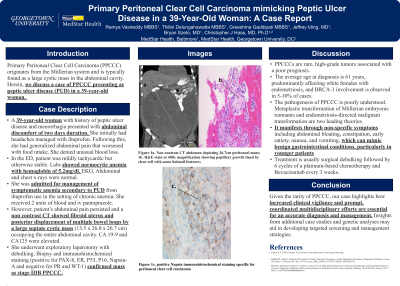
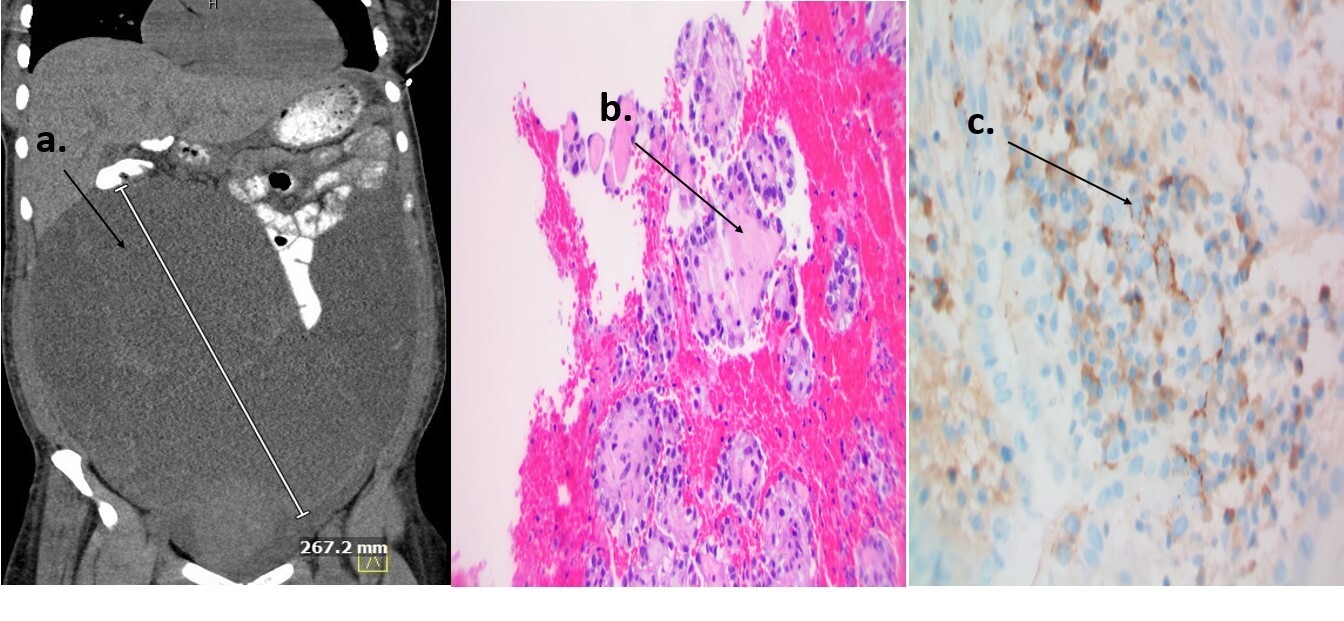
Case Description/Methods: A 39-year-old woman with menorrhagia and peptic ulcer disease presented with abdominal discomfort of 2 days duration. She initially had headaches managed with ibuprofen. Following this, she had generalized abdominal pain with bloating that worsened with food, and no relief with use of stool softeners. She had associated dizziness with palpitations, chest pressure and exertional dyspnea. In the emergency department, the patient was mildly tachycardic but otherwise stable. On exam, she had a distended abdomen with generalized tenderness and normoactive bowel sounds. Labs showed normocytic anemia with a hemoglobin of 5.2 mg/dL. EKG, Abdominal and chest x-rays were normal. A non-contrast CT of abdomen and pelvis showed a fibroid uterus and posterior displacement of multiple bowel loops by a large septate cystic mass (13.5 x 26.0 x 26.7 cm) occupying the entire abdominal cavity. Elevated CA125 and CA 19-9 were also noted. She underwent exploratory laparotomy with mass resection, partial omentectomy, left colectomy (given extension into transverse colon), appendectomy and total abdominal hysterectomy with bilateral salpingectomy. Biopsy and immunohistochemical staining (positive for PAX-8, ER, P53, P16, Napsin-A and negative for PR and WT-1) confirmed mass as stage IIIB PPCCC. There was no evidence of malignancy or endometriotic foci in other tissue samples. The patient was discharged with a plan for outpatient platinum-based chemotherapy and genetic counselling.
Discussion: Given the rarity of PPCCC, our case highlights how increased clinical vigilance and prompt, coordinated multidisciplinary efforts are essential for an accurate diagnosis. Currently, there are no established management guidelines, however, initial treatment with surgical debulking followed by chemotherapy is often practiced. Further insights from case studies and genetic analyses may help in developing targeted screening and management strategies.

Disclosures:
Ramya Vasireddy, MBBS1, Thilini Delungahawatta, MD2, Greeshma Gaddipati, MBBS1, Jeffrey Iding, MD3, Bryan Szeto, MD3, Christopher Haas, MD, PhD2. P3288 - Primary Peritoneal Clear Cell Carcinoma in a 39-Year-Old Woman: A Case Report, ACG 2024 Annual Scientific Meeting Abstracts. Philadelphia, PA: American College of Gastroenterology.
1MedStar Union Memorial Hospital, Baltimore, MD; 2MedStar Health, Baltimore, MD; 3MedStar Franklin Square Medical Center, Baltimore, MD
Introduction: Primary peritoneal clear cell carcinoma (PPCCC) is a rare abdominal tumor with a reported incidence of 7 cases per million persons. The average age at diagnosis is 61 years, predominantly affecting white females with endometriosis, and BRCA-1 involvement is observed in 5-10% of cases. Pathogenesis remains unclear but is thought to arise from remnant ovarian tissue in peritoneum following embryonic migration. Patients usually present with nonspecific symptoms associated with tumor mass effect, often conferring diagnostic delay and poor prognosis.
Case Description/Methods: A 39-year-old woman with menorrhagia and peptic ulcer disease presented with abdominal discomfort of 2 days duration. She initially had headaches managed with ibuprofen. Following this, she had generalized abdominal pain with bloating that worsened with food, and no relief with use of stool softeners. She had associated dizziness with palpitations, chest pressure and exertional dyspnea. In the emergency department, the patient was mildly tachycardic but otherwise stable. On exam, she had a distended abdomen with generalized tenderness and normoactive bowel sounds. Labs showed normocytic anemia with a hemoglobin of 5.2 mg/dL. EKG, Abdominal and chest x-rays were normal. A non-contrast CT of abdomen and pelvis showed a fibroid uterus and posterior displacement of multiple bowel loops by a large septate cystic mass (13.5 x 26.0 x 26.7 cm) occupying the entire abdominal cavity. Elevated CA125 and CA 19-9 were also noted. She underwent exploratory laparotomy with mass resection, partial omentectomy, left colectomy (given extension into transverse colon), appendectomy and total abdominal hysterectomy with bilateral salpingectomy. Biopsy and immunohistochemical staining (positive for PAX-8, ER, P53, P16, Napsin-A and negative for PR and WT-1) confirmed mass as stage IIIB PPCCC. There was no evidence of malignancy or endometriotic foci in other tissue samples. The patient was discharged with a plan for outpatient platinum-based chemotherapy and genetic counselling.
Discussion: Given the rarity of PPCCC, our case highlights how increased clinical vigilance and prompt, coordinated multidisciplinary efforts are essential for an accurate diagnosis. Currently, there are no established management guidelines, however, initial treatment with surgical debulking followed by chemotherapy is often practiced. Further insights from case studies and genetic analyses may help in developing targeted screening and management strategies.

Figure: Figure 1: a. Non-contrast CT abdomen depicting 26.7cm peritoneal mass; b. H&E stain at 400x magnification showing papillary growth lined by clear cell with some hobnail features; c. positive Napsin immunohistochemical staining specific for peritoneal clear cell carcinoma
Disclosures:
Ramya Vasireddy indicated no relevant financial relationships.
Thilini Delungahawatta indicated no relevant financial relationships.
Greeshma Gaddipati indicated no relevant financial relationships.
Jeffrey Iding indicated no relevant financial relationships.
Bryan Szeto indicated no relevant financial relationships.
Christopher Haas indicated no relevant financial relationships.
Ramya Vasireddy, MBBS1, Thilini Delungahawatta, MD2, Greeshma Gaddipati, MBBS1, Jeffrey Iding, MD3, Bryan Szeto, MD3, Christopher Haas, MD, PhD2. P3288 - Primary Peritoneal Clear Cell Carcinoma in a 39-Year-Old Woman: A Case Report, ACG 2024 Annual Scientific Meeting Abstracts. Philadelphia, PA: American College of Gastroenterology.
