Monday Poster Session
Category: Small Intestine
P3291 - A Rare Case of Segmental Ileal Dilatation Causing Severe Malnutrition in a Young Woman
Monday, October 28, 2024
10:30 AM - 4:00 PM ET
Location: Exhibit Hall E

Has Audio

Shalva Eliava, MD
Englewood Hospital and Medical Center
Englewood, NJ
Presenting Author(s)
Award: Presidential Poster Award
Shalva Eliava, MD1, Danielle Pope, MD2, Dipal Patel, MD1, Mitchell Spinnell, MD1
1Englewood Hospital and Medical Center, Englewood, NJ; 2Englewood Hospital and Medical Center, Bogota, NJ
Introduction: Segmental intestinal dilatation (SID) is a rare gastrointestinal (GI) disorder characterized by a markedly dilated segment flanked by normal-caliber afferent and efferent bowel. The majority of SID cases have been reported in neonates and infants. Patients may present with GI bleeding, abdominal pain, anemia, failure to thrive, and signs of intestinal obstruction. We present an extremely rare case of segmental ileal dilatation in a young woman.
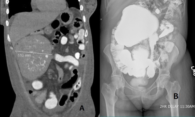
Case Description/Methods: A 44-year-old female with a history of lactose intolerance presented with anasarca, intermittent watery diarrhea, abdominal distention, and 20-pound weight loss for three months. Laboratory testing showed severe albumin deficiency, low vitamin D level, and normocytic anemia. CT abdomen revealed focal marked dilatation of distal ileum measuring up to 11 cm with abrupt caliber change without significant upstream dilatation, suggesting segmental mega ileum vs ileal dysgenesis.
Extensive workup for diarrhea and malnutrition showed normal 24-hour urine protein and 24-hour stool Alpha-1-Antitrypsin clearance. Testing for TSH, C-reactive protein, erythrocyte sedimentation rate, ova and parasite, clostridium difficile, HIV, celiac disease, and GI pathogen panel returned negative. Colonoscopy and esophagogastroduodenoscopy with small bowel biopsy were unrevealing. Fecal pancreatic elastase was 38, consistent with severe pancreatic insufficiency. Surgery was consulted for resection of a dilated ileal segment, but given her poor nutritional status and high risk for surgical complications, she was commenced on total parenteral nutrition and discharged home with close follow-up for surgical planning.
Discussion: SID is a rare congenital abnormality and is scarcely described in adults. It may be associated with Meckel’s diverticulum and omphalocele. Dilated segments could contain ectopic tissue, including gastric, pancreatic, and esophageal histology. Classic CT scan findings are sharply demarcated segmental bowel dilatation with or without air-fluid levels.
Our unique case demonstrates that the most common presenting symptom, GI bleeding, is not always present. Instead, our patient presented with severe malnutrition, abdominal pain, and signs of bowel obstruction. The cause of severe malnutrition is not entirely clear. Likely, it was a combination of pancreatic insufficiency, poor oral intake, and malabsorption due to a dilated ileum.
Definitive treatment of SID is surgical resection of dilated segments with excellent long-term prognosis.

Disclosures:
Shalva Eliava, MD1, Danielle Pope, MD2, Dipal Patel, MD1, Mitchell Spinnell, MD1. P3291 - A Rare Case of Segmental Ileal Dilatation Causing Severe Malnutrition in a Young Woman, ACG 2024 Annual Scientific Meeting Abstracts. Philadelphia, PA: American College of Gastroenterology.
Shalva Eliava, MD1, Danielle Pope, MD2, Dipal Patel, MD1, Mitchell Spinnell, MD1
1Englewood Hospital and Medical Center, Englewood, NJ; 2Englewood Hospital and Medical Center, Bogota, NJ
Introduction: Segmental intestinal dilatation (SID) is a rare gastrointestinal (GI) disorder characterized by a markedly dilated segment flanked by normal-caliber afferent and efferent bowel. The majority of SID cases have been reported in neonates and infants. Patients may present with GI bleeding, abdominal pain, anemia, failure to thrive, and signs of intestinal obstruction. We present an extremely rare case of segmental ileal dilatation in a young woman.
Case Description/Methods: A 44-year-old female with a history of lactose intolerance presented with anasarca, intermittent watery diarrhea, abdominal distention, and 20-pound weight loss for three months. Laboratory testing showed severe albumin deficiency, low vitamin D level, and normocytic anemia. CT abdomen revealed focal marked dilatation of distal ileum measuring up to 11 cm with abrupt caliber change without significant upstream dilatation, suggesting segmental mega ileum vs ileal dysgenesis.
Extensive workup for diarrhea and malnutrition showed normal 24-hour urine protein and 24-hour stool Alpha-1-Antitrypsin clearance. Testing for TSH, C-reactive protein, erythrocyte sedimentation rate, ova and parasite, clostridium difficile, HIV, celiac disease, and GI pathogen panel returned negative. Colonoscopy and esophagogastroduodenoscopy with small bowel biopsy were unrevealing. Fecal pancreatic elastase was 38, consistent with severe pancreatic insufficiency. Surgery was consulted for resection of a dilated ileal segment, but given her poor nutritional status and high risk for surgical complications, she was commenced on total parenteral nutrition and discharged home with close follow-up for surgical planning.
Discussion: SID is a rare congenital abnormality and is scarcely described in adults. It may be associated with Meckel’s diverticulum and omphalocele. Dilated segments could contain ectopic tissue, including gastric, pancreatic, and esophageal histology. Classic CT scan findings are sharply demarcated segmental bowel dilatation with or without air-fluid levels.
Our unique case demonstrates that the most common presenting symptom, GI bleeding, is not always present. Instead, our patient presented with severe malnutrition, abdominal pain, and signs of bowel obstruction. The cause of severe malnutrition is not entirely clear. Likely, it was a combination of pancreatic insufficiency, poor oral intake, and malabsorption due to a dilated ileum.
Definitive treatment of SID is surgical resection of dilated segments with excellent long-term prognosis.

Figure: Image A: Coronal CT scan view showing focal marked dilatation involving a loop of distal ileum in the right hemiabdomen up to 11 cm with abrupt caliber change without significant upstream dilation.
Image B : AP X-Ray small bowel series showing marked dilation of the loop of ileum in the right hemiabdomen without evidence of obstruction or delayed transit.
Image B : AP X-Ray small bowel series showing marked dilation of the loop of ileum in the right hemiabdomen without evidence of obstruction or delayed transit.
Disclosures:
Shalva Eliava indicated no relevant financial relationships.
Danielle Pope indicated no relevant financial relationships.
Dipal Patel indicated no relevant financial relationships.
Mitchell Spinnell indicated no relevant financial relationships.
Shalva Eliava, MD1, Danielle Pope, MD2, Dipal Patel, MD1, Mitchell Spinnell, MD1. P3291 - A Rare Case of Segmental Ileal Dilatation Causing Severe Malnutrition in a Young Woman, ACG 2024 Annual Scientific Meeting Abstracts. Philadelphia, PA: American College of Gastroenterology.


