Monday Poster Session
Category: Small Intestine
P3306 - Terminal Ileitis in a Young Adult: Unusual Presentation of IgA Vasculitis
Monday, October 28, 2024
10:30 AM - 4:00 PM ET
Location: Exhibit Hall E

Has Audio

Osama Alshakhatreh, MD
Albany Medical Center
Albany, NY
Presenting Author(s)
Osama Alshakhatreh, MD, Ahmad Abulawi, MD, Khaled Elsokary, DO, Raya Alashram, MD, Hasan B. Aydin, MD, Hubert H Nietsch, MD, Seth Richter, MD
Albany Medical Center, Albany, NY
Introduction: IgA vasculitis, also known as Henoch-Schönlein purpura (HSP), is an immune-mediated vasculitis affecting small vessels across various organs. While terminal ileitis (TI) in young adults usually suggests Crohn’s disease, it is crucial to consider other rare etiologies, especially when accompanied by a rash. Among these, vasculitis and infections warrant attention. Here, we present a case of terminal ileitis in a young female diagnosed with IgA vasculitis.
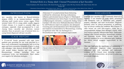
Case Description/Methods: A 25-year-old female presented with right lower quadrant pain persisting for one week preceded by a three-week erythematous maculopapular rash involving upper and lower extremities bilaterally (Figure 1), along with arthralgia. Labs showed elevated ESR, and CRP with normal kidney function and urine analysis. Rheumatologic workup, including ANA and complement levels, was unremarkable. Stool studies were negative for infectious etiologies. Abdominal CT revealed terminal ileum thickening, confirmed by colonoscopy showing erythematous, inflamed, and ulcerated mucosa (Figure 2). Pathological analysis confirmed acute ileitis (Figure 3) and skin biopsies yielded spongiotic dermatitis, a nonspecific finding. Initial treatment with intravenous Methylprednisolone followed by oral Prednisone improved symptoms, but subsequent worsening pain and hematochezia led to Dexamethasone and Anakinra recommendation by Rheumatology, resulting in significant relief. She was discharged on a Dexamethasone taper with continued improvement.
Discussion: Although IgA vasculitis is more commonly observed in children, it can manifest in young adults, presenting with symptoms such as abdominal pain, palpable purpura, arthralgia, and renal involvement. Diagnosis primarily relies on clinical presentation, as pathology may not always confirm the condition. Skin biopsies of older lesions can yield false-negative results due to immunoglobulin leakage from damaged vessels, while ileal biopsies typically indicate acute ileitis. Endoscopic findings often include petechiae, hemorrhagic erosions, and hyperemic lesions. While steroids remain the mainstay treatment for severe abdominal pain, Anakinra has shown partial symptom improvement in the literature.
This case highlights the significance of maintaining a broad differential diagnosis when encountering abdominal pain with radiologic and endoscopic evidence of TI. Clinical clues, including symptom acuity and rash characteristics, are pivotal in achieving an accurate diagnosis.

Disclosures:
Osama Alshakhatreh, MD, Ahmad Abulawi, MD, Khaled Elsokary, DO, Raya Alashram, MD, Hasan B. Aydin, MD, Hubert H Nietsch, MD, Seth Richter, MD. P3306 - Terminal Ileitis in a Young Adult: Unusual Presentation of IgA Vasculitis, ACG 2024 Annual Scientific Meeting Abstracts. Philadelphia, PA: American College of Gastroenterology.
Albany Medical Center, Albany, NY
Introduction: IgA vasculitis, also known as Henoch-Schönlein purpura (HSP), is an immune-mediated vasculitis affecting small vessels across various organs. While terminal ileitis (TI) in young adults usually suggests Crohn’s disease, it is crucial to consider other rare etiologies, especially when accompanied by a rash. Among these, vasculitis and infections warrant attention. Here, we present a case of terminal ileitis in a young female diagnosed with IgA vasculitis.
Case Description/Methods: A 25-year-old female presented with right lower quadrant pain persisting for one week preceded by a three-week erythematous maculopapular rash involving upper and lower extremities bilaterally (Figure 1), along with arthralgia. Labs showed elevated ESR, and CRP with normal kidney function and urine analysis. Rheumatologic workup, including ANA and complement levels, was unremarkable. Stool studies were negative for infectious etiologies. Abdominal CT revealed terminal ileum thickening, confirmed by colonoscopy showing erythematous, inflamed, and ulcerated mucosa (Figure 2). Pathological analysis confirmed acute ileitis (Figure 3) and skin biopsies yielded spongiotic dermatitis, a nonspecific finding. Initial treatment with intravenous Methylprednisolone followed by oral Prednisone improved symptoms, but subsequent worsening pain and hematochezia led to Dexamethasone and Anakinra recommendation by Rheumatology, resulting in significant relief. She was discharged on a Dexamethasone taper with continued improvement.
Discussion: Although IgA vasculitis is more commonly observed in children, it can manifest in young adults, presenting with symptoms such as abdominal pain, palpable purpura, arthralgia, and renal involvement. Diagnosis primarily relies on clinical presentation, as pathology may not always confirm the condition. Skin biopsies of older lesions can yield false-negative results due to immunoglobulin leakage from damaged vessels, while ileal biopsies typically indicate acute ileitis. Endoscopic findings often include petechiae, hemorrhagic erosions, and hyperemic lesions. While steroids remain the mainstay treatment for severe abdominal pain, Anakinra has shown partial symptom improvement in the literature.
This case highlights the significance of maintaining a broad differential diagnosis when encountering abdominal pain with radiologic and endoscopic evidence of TI. Clinical clues, including symptom acuity and rash characteristics, are pivotal in achieving an accurate diagnosis.

Figure: Figure (A) Extensive erythematous rash involving lower extremities. Figure (B) Congested, erythematous, inflamed mucosa in the distal ileum. Figure (C) Terminal ileal mucosa with acute inflammation.
Disclosures:
Osama Alshakhatreh indicated no relevant financial relationships.
Ahmad Abulawi indicated no relevant financial relationships.
Khaled Elsokary indicated no relevant financial relationships.
Raya Alashram indicated no relevant financial relationships.
Hasan Aydin indicated no relevant financial relationships.
Hubert H Nietsch indicated no relevant financial relationships.
Seth Richter indicated no relevant financial relationships.
Osama Alshakhatreh, MD, Ahmad Abulawi, MD, Khaled Elsokary, DO, Raya Alashram, MD, Hasan B. Aydin, MD, Hubert H Nietsch, MD, Seth Richter, MD. P3306 - Terminal Ileitis in a Young Adult: Unusual Presentation of IgA Vasculitis, ACG 2024 Annual Scientific Meeting Abstracts. Philadelphia, PA: American College of Gastroenterology.
