Monday Poster Session
Category: Stomach
P3344 - A Rare Case of Primary Gastric Small Cell Carcinoma
Monday, October 28, 2024
10:30 AM - 4:00 PM ET
Location: Exhibit Hall E

- PM
Pujyitha Mandiga, MD
Ascension Macomb-Oakland Hospital
Parker, CO
Presenting Author(s)
Pujyitha Mandiga, MD1, Saketh Mandiga, MBBS2, Manthan Patel, MD3, Mohammed Barawi, MD4
1Ascension Macomb-Oakland Hospital, Parker, CO; 2Osmania General Hospital and Medical College, Hyderabad, Telangana, India; 3Ascension Macomb-Oakland Hospital, Muskegon, MI; 4Ascension St. John Hospital, Detroit, MI
Introduction: Small cell carcinoma (SmCC), is a highly uncommon and aggressive histological subtype of gastric cancer that poses significant challenges in diagnosis and management. In the literature review, only few cases of SmCC were reported in the Genitourinary system, larynx, salivary glands.
Gastric SmCC accounts for only 0.1% of all gastric carcinomas. They originate from neuroendocrine APUD cells. Among all the extrapulmonary SmCC, gastrointestinal SmCC carries the worst prognosis. It tends to metastasize widely, median survival was reported at about one year.
Case Description/Methods: Gastric SmCC accounts for only 0.1% of all gastric carcinomas. They originate from neuroendocrine APUD cells and tends to metastasize widely, median survival was reported at about one year. The combination of chemotherapy is the mainstay of treatment, surgery and radiation would significantly improve survival but with poor outcomes.
Discussion: We are presenting a case of a 70 year old male with a Hx HTN, HLD presents with persistent epigastric pain, burning sensation, and fatigue over 8 weeks. Despite using H2RAs, PPIs, carafate, his symptoms persisted, which warranted for further investigation.
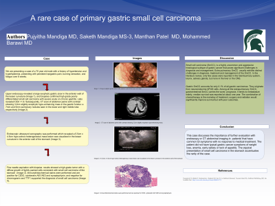
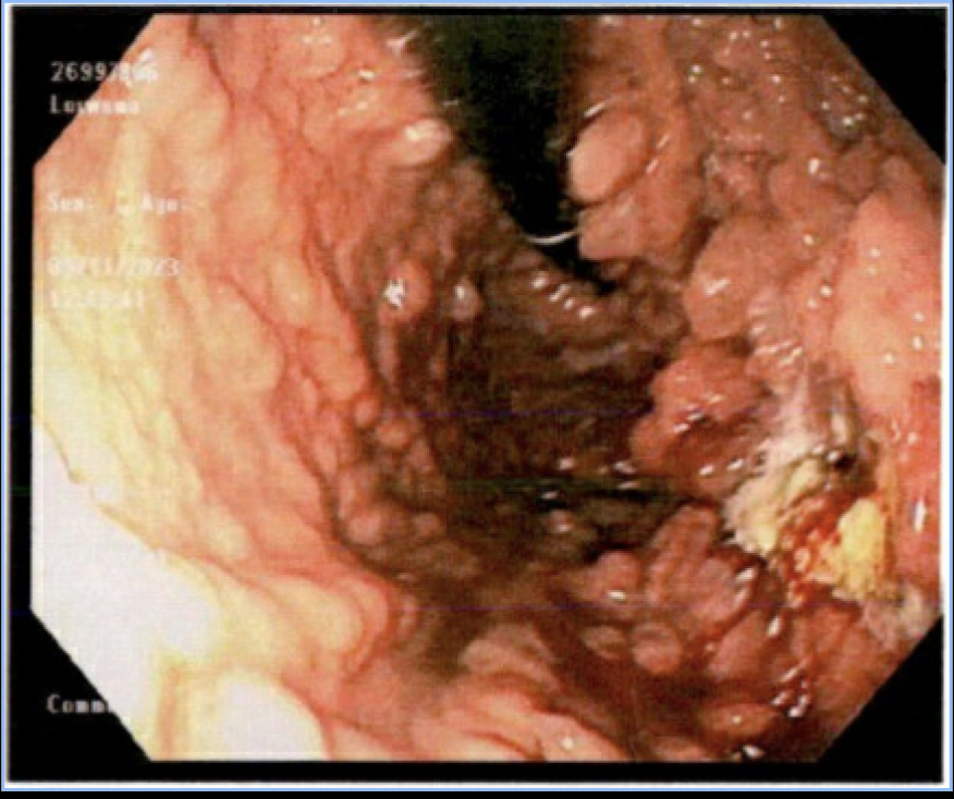
EGD revealed a large exophytic gastric ulcer in the anterior wall of the lesser curvature (Image 1), and biopsies confirmed high grade poorly differentiated SmCC with severe acute on chronic gastritis. CEA < 1.8. Subsequently, CT scan of abdomen pelvis with contrast showing 3.2cm slightly exophytic hypo-enhancing mass in the gastric fundus and multiple nodules in the lungs.
EUS was performed which revealed a 5.5cm x 4.5cm hypo-echoic heterogeneous mass lesion was visualized in the lesser curvature in the anterior wall of the stomach. FNA with biopsies results showed a high grade tumor with a diffuse growth of tightly packed cells consistent with SmCC of the stomach. Immunohistochemical stains were performed and are positive for CDX2, cytokeratin AE1/AE3 and synaptophysin, and negative for chromogranin and TTF1 supported the diagnosis of SmCC. Further imaging did not reveal any metastasis. Patient was started on chemotherapy.
This case discusses the importance of further investigation in patients' that have common GI symptoms with no response to medical treatment. The patient did not have typical gastric cancer symptoms of weight loss, anemia, early satiety or lack of appetite. The atypical presentation of small cell carcinoma in the stomach accentuates the rarity of the case.

Disclosures:
Pujyitha Mandiga, MD1, Saketh Mandiga, MBBS2, Manthan Patel, MD3, Mohammed Barawi, MD4. P3344 - A Rare Case of Primary Gastric Small Cell Carcinoma, ACG 2024 Annual Scientific Meeting Abstracts. Philadelphia, PA: American College of Gastroenterology.
1Ascension Macomb-Oakland Hospital, Parker, CO; 2Osmania General Hospital and Medical College, Hyderabad, Telangana, India; 3Ascension Macomb-Oakland Hospital, Muskegon, MI; 4Ascension St. John Hospital, Detroit, MI
Introduction: Small cell carcinoma (SmCC), is a highly uncommon and aggressive histological subtype of gastric cancer that poses significant challenges in diagnosis and management. In the literature review, only few cases of SmCC were reported in the Genitourinary system, larynx, salivary glands.
Gastric SmCC accounts for only 0.1% of all gastric carcinomas. They originate from neuroendocrine APUD cells. Among all the extrapulmonary SmCC, gastrointestinal SmCC carries the worst prognosis. It tends to metastasize widely, median survival was reported at about one year.
Case Description/Methods: Gastric SmCC accounts for only 0.1% of all gastric carcinomas. They originate from neuroendocrine APUD cells and tends to metastasize widely, median survival was reported at about one year. The combination of chemotherapy is the mainstay of treatment, surgery and radiation would significantly improve survival but with poor outcomes.
Discussion: We are presenting a case of a 70 year old male with a Hx HTN, HLD presents with persistent epigastric pain, burning sensation, and fatigue over 8 weeks. Despite using H2RAs, PPIs, carafate, his symptoms persisted, which warranted for further investigation.
EGD revealed a large exophytic gastric ulcer in the anterior wall of the lesser curvature (Image 1), and biopsies confirmed high grade poorly differentiated SmCC with severe acute on chronic gastritis. CEA < 1.8. Subsequently, CT scan of abdomen pelvis with contrast showing 3.2cm slightly exophytic hypo-enhancing mass in the gastric fundus and multiple nodules in the lungs.
EUS was performed which revealed a 5.5cm x 4.5cm hypo-echoic heterogeneous mass lesion was visualized in the lesser curvature in the anterior wall of the stomach. FNA with biopsies results showed a high grade tumor with a diffuse growth of tightly packed cells consistent with SmCC of the stomach. Immunohistochemical stains were performed and are positive for CDX2, cytokeratin AE1/AE3 and synaptophysin, and negative for chromogranin and TTF1 supported the diagnosis of SmCC. Further imaging did not reveal any metastasis. Patient was started on chemotherapy.
This case discusses the importance of further investigation in patients' that have common GI symptoms with no response to medical treatment. The patient did not have typical gastric cancer symptoms of weight loss, anemia, early satiety or lack of appetite. The atypical presentation of small cell carcinoma in the stomach accentuates the rarity of the case.

Figure: EGD- Mass in the lesser curvature in the anterior wall of the stomach
Disclosures:
Pujyitha Mandiga indicated no relevant financial relationships.
Saketh Mandiga indicated no relevant financial relationships.
Manthan Patel indicated no relevant financial relationships.
Mohammed Barawi: Abbvie – Speakers Bureau. Boston scietific – Consultant. Gilead – Speakers Bureau. Olympus – Consultant.
Pujyitha Mandiga, MD1, Saketh Mandiga, MBBS2, Manthan Patel, MD3, Mohammed Barawi, MD4. P3344 - A Rare Case of Primary Gastric Small Cell Carcinoma, ACG 2024 Annual Scientific Meeting Abstracts. Philadelphia, PA: American College of Gastroenterology.
