Monday Poster Session
Category: Stomach
P3379 - A Rare Case of Primary Gastric Actinomycosis Associated With Gastric Adenocarcinoma and Partial Gatsrectomy
Monday, October 28, 2024
10:30 AM - 4:00 PM ET
Location: Exhibit Hall E

Has Audio

Annie Shergill, MD
Larkin Community Hospital
Miami Beach, FL
Presenting Author(s)
Annie Shergill, MD1, Jose Proenza, MD2
1Larkin Community Hospital, Miami Beach, FL; 2West Palm Beach VA Medical Center, West Palm beach, FL
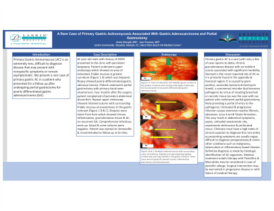
Introduction: Primary Gastric Actinomycosis (AC) is an extremely rare, difficult to diagnose disease that may present with nonspecific symptoms or remain asymptomatic. We present a rare case of primary gastric AC in a patient who presented for a follow up after undergoing partial gastrectomy for poorly differentiated gastric adenocarcinoma (GA).
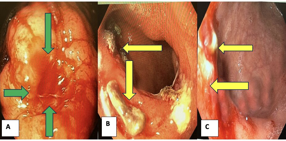
Case Description/Methods: 65 year-old male with history of GERD presented to the clinic with persistent dyspepsia. Patient underwent upper endoscopy which showed an area of indurated, friable mucosa in greater curvature (Figure 1 A) which was biopsied. Biopsy showed poorly differentiated gastric adenocarcinoma. Patient underwent partial gastrectomy with primary hand sewn anastomosis. Four months after the surgery patient complained of persistent abdominal discomfort. Repeat upper endoscopy showed retained sutures with surrounding friable mucosa at anastomosis in the gastric remnant (Figure 1 B & C). Biopsies were taken from here which showed chronic inflammation, granulomatous tissue & AC, no recurrent GA. Comprehensive infectious work up- blood & urine cultures were negative. Patient was started on Amoxicillin & recommended to follow up in GI clinic.
Discussion: Primary gastric AC is a rare (with only a few of case reports to date), chronic granulomatous disease with an indolent course associated with significant morbidity. Stomach is the rarest reported site of AC as it is primarily found in the appendix & ileocecal region. It is caused by gram positive, anaerobic bacteria Actonimyces Israelii, a commensal microbe that becomes pathogenic by virtue of invading breached or necrotic tissue (as was the case with our patient who underwent partial gastrectomy, likely providing a portal of entry to the pathogens). Untreated & progressive infection causes extensive reactive fibrosis, abscesses, sinus tract & fistula formation. This may result in abdominal symptoms, sepsis, unhealed anastomotic site, anastomotic dehiscence & perforated viscus. Clinicians must have a high index of clinical suspicion to diagnose this rare entity as presenting symptoms are usually vague, difficult to diagnose preoperatively & mimic other conditions such as malignancy, tuberculosis or inflammatory bowel disease. Definitive diagnosis is made by histologic identification of AC organisms. Medical treatment entails therapy with Penicillins & Macrolides may be considered in case of penicillin allergy. Surgical intervention may be warranted in progressive disease or with failure of medical therapy.

Disclosures:
Annie Shergill, MD1, Jose Proenza, MD2. P3379 - A Rare Case of Primary Gastric Actinomycosis Associated With Gastric Adenocarcinoma and Partial Gatsrectomy, ACG 2024 Annual Scientific Meeting Abstracts. Philadelphia, PA: American College of Gastroenterology.
1Larkin Community Hospital, Miami Beach, FL; 2West Palm Beach VA Medical Center, West Palm beach, FL
Introduction: Primary Gastric Actinomycosis (AC) is an extremely rare, difficult to diagnose disease that may present with nonspecific symptoms or remain asymptomatic. We present a rare case of primary gastric AC in a patient who presented for a follow up after undergoing partial gastrectomy for poorly differentiated gastric adenocarcinoma (GA).
Case Description/Methods: 65 year-old male with history of GERD presented to the clinic with persistent dyspepsia. Patient underwent upper endoscopy which showed an area of indurated, friable mucosa in greater curvature (Figure 1 A) which was biopsied. Biopsy showed poorly differentiated gastric adenocarcinoma. Patient underwent partial gastrectomy with primary hand sewn anastomosis. Four months after the surgery patient complained of persistent abdominal discomfort. Repeat upper endoscopy showed retained sutures with surrounding friable mucosa at anastomosis in the gastric remnant (Figure 1 B & C). Biopsies were taken from here which showed chronic inflammation, granulomatous tissue & AC, no recurrent GA. Comprehensive infectious work up- blood & urine cultures were negative. Patient was started on Amoxicillin & recommended to follow up in GI clinic.
Discussion: Primary gastric AC is a rare (with only a few of case reports to date), chronic granulomatous disease with an indolent course associated with significant morbidity. Stomach is the rarest reported site of AC as it is primarily found in the appendix & ileocecal region. It is caused by gram positive, anaerobic bacteria Actonimyces Israelii, a commensal microbe that becomes pathogenic by virtue of invading breached or necrotic tissue (as was the case with our patient who underwent partial gastrectomy, likely providing a portal of entry to the pathogens). Untreated & progressive infection causes extensive reactive fibrosis, abscesses, sinus tract & fistula formation. This may result in abdominal symptoms, sepsis, unhealed anastomotic site, anastomotic dehiscence & perforated viscus. Clinicians must have a high index of clinical suspicion to diagnose this rare entity as presenting symptoms are usually vague, difficult to diagnose preoperatively & mimic other conditions such as malignancy, tuberculosis or inflammatory bowel disease. Definitive diagnosis is made by histologic identification of AC organisms. Medical treatment entails therapy with Penicillins & Macrolides may be considered in case of penicillin allergy. Surgical intervention may be warranted in progressive disease or with failure of medical therapy.

Figure: Figure 1 A: Area of induration and friability (green arrows) in the greater curvature seen during initial upper endoscopy, this was biopsied to be poorly differentiated gastric adenocarcinoma. Figure 1 B & C: Multiple retained sutures with surrounding areas of erythema, friability and some bleeding (yellow arrows) seen post-operatively in the gastric remnant. These areas were biopsied & showed chronic inflammation, granulomas and actinomycosis.
Disclosures:
Annie Shergill indicated no relevant financial relationships.
Jose Proenza indicated no relevant financial relationships.
Annie Shergill, MD1, Jose Proenza, MD2. P3379 - A Rare Case of Primary Gastric Actinomycosis Associated With Gastric Adenocarcinoma and Partial Gatsrectomy, ACG 2024 Annual Scientific Meeting Abstracts. Philadelphia, PA: American College of Gastroenterology.
