Monday Poster Session
Category: Stomach
P3400 - GIST Tumor Presenting as Asymptomatic Anemia: A Case Report
Monday, October 28, 2024
10:30 AM - 4:00 PM ET
Location: Exhibit Hall E

Has Audio

Nosheen Hafeez, MD
Baptist Health-University of Arkansas for Medical Sciences
North Little Rock, AR
Presenting Author(s)
Nosheen Hafeez, MD1, Fatima Khalid, MD2, Buraq Al-Ghaieb, MD1, Ekta Singh, MD1, Ahmad Abdulmajid, MD1
1Baptist Health-University of Arkansas for Medical Sciences, North Little Rock, AR; 2Baptist Health-UAMS, Overland Park, KS
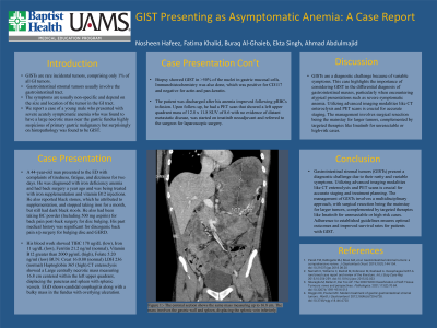
Introduction: GISTs are rare incidental tumors, comprising only 1% of all GI tumors. we report a case of a young male who presented with severe symptomatic anemia who was found to have a large necrotic mass near the gastric fundus highly suspicious of primary gastric malignancy but surprisingly on histopathology was found to be GIST.
Case Description/Methods: A 44-year-old man presented to the ED with complaints of tiredness, fatigue, and dizziness for two days. He was diagnosed with iron deficiency anemia and had back surgery a year ago and was being treated with iron supplementation and vitamin B12 injections. He also reported black stones, which he attributed to supplementation, and stopped taking iron for a month, but still had dark black stools. He also had been taking BC powder (Including 500 mg aspirin) for back pain post-back surgery for disc bulging. His past medical history was significant for discogenic back pain s/p surgery for bulging disc and GERD.
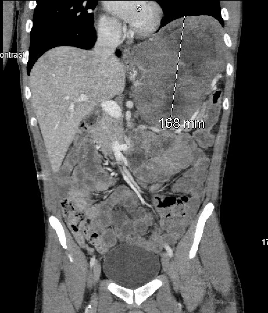
His blood work showed TIBC 178 ug/dL (low), Iron 11 ug/dL (low), Ferritin 21.2 ng/ml (normal), Vitamin B12 greater than 2000 pg/mL (high), Folate 5.20 ng/ml (low) BUN: Creat 16:0.80 (normal) LDH 236 (normal) Haptoglobin 365 (high).CT enteroclysis showed a Large centrally necrotic mass measuring 16.8 cm centered within the left upper quadrant, displacing the pancreas and spleen with splenic vessels. EGD shows candidal esophagitis along with a bulky mass in the fundus with overlying ulceration.
Biopsy showed GIST in >50% of the nuclei in gastric mucosal cells. Immunohistochemistry was also done, which was positive for CD117 and negative for actin and pan-keratin. The patient was discharged after his anemia improved following pRBCs infusion. Upon follow-up, he had a PET scan that showed a left upper quadrant mass of 12.8 x 11.8 SUV of 8.6 with no evidence of distant metastatic disease, was started on imatinib neoadjuvant and referred to the surgeon for laparoscopic surgery.
Discussion: GISTs are a diagnostic challenge because of variable symptoms. This case highlights the importance of considering GIST in the differential diagnosis of gastrointestinal masses, particularly when encountering atypical presentations such as severe symptomatic anemia. Utilizing advanced imaging modalities like CT enteroclysis and PET scans is crucial for accurate staging. The management involves surgical resection being the mainstay for larger tumors, complemented by targeted therapies like Imatinib for unresectable or high-risk cases.

Disclosures:
Nosheen Hafeez, MD1, Fatima Khalid, MD2, Buraq Al-Ghaieb, MD1, Ekta Singh, MD1, Ahmad Abdulmajid, MD1. P3400 - GIST Tumor Presenting as Asymptomatic Anemia: A Case Report, ACG 2024 Annual Scientific Meeting Abstracts. Philadelphia, PA: American College of Gastroenterology.
1Baptist Health-University of Arkansas for Medical Sciences, North Little Rock, AR; 2Baptist Health-UAMS, Overland Park, KS
Introduction: GISTs are rare incidental tumors, comprising only 1% of all GI tumors. we report a case of a young male who presented with severe symptomatic anemia who was found to have a large necrotic mass near the gastric fundus highly suspicious of primary gastric malignancy but surprisingly on histopathology was found to be GIST.
Case Description/Methods: A 44-year-old man presented to the ED with complaints of tiredness, fatigue, and dizziness for two days. He was diagnosed with iron deficiency anemia and had back surgery a year ago and was being treated with iron supplementation and vitamin B12 injections. He also reported black stones, which he attributed to supplementation, and stopped taking iron for a month, but still had dark black stools. He also had been taking BC powder (Including 500 mg aspirin) for back pain post-back surgery for disc bulging. His past medical history was significant for discogenic back pain s/p surgery for bulging disc and GERD.
His blood work showed TIBC 178 ug/dL (low), Iron 11 ug/dL (low), Ferritin 21.2 ng/ml (normal), Vitamin B12 greater than 2000 pg/mL (high), Folate 5.20 ng/ml (low) BUN: Creat 16:0.80 (normal) LDH 236 (normal) Haptoglobin 365 (high).CT enteroclysis showed a Large centrally necrotic mass measuring 16.8 cm centered within the left upper quadrant, displacing the pancreas and spleen with splenic vessels. EGD shows candidal esophagitis along with a bulky mass in the fundus with overlying ulceration.
Biopsy showed GIST in >50% of the nuclei in gastric mucosal cells. Immunohistochemistry was also done, which was positive for CD117 and negative for actin and pan-keratin. The patient was discharged after his anemia improved following pRBCs infusion. Upon follow-up, he had a PET scan that showed a left upper quadrant mass of 12.8 x 11.8 SUV of 8.6 with no evidence of distant metastatic disease, was started on imatinib neoadjuvant and referred to the surgeon for laparoscopic surgery.
Discussion: GISTs are a diagnostic challenge because of variable symptoms. This case highlights the importance of considering GIST in the differential diagnosis of gastrointestinal masses, particularly when encountering atypical presentations such as severe symptomatic anemia. Utilizing advanced imaging modalities like CT enteroclysis and PET scans is crucial for accurate staging. The management involves surgical resection being the mainstay for larger tumors, complemented by targeted therapies like Imatinib for unresectable or high-risk cases.

Figure: Figure 1:- The coronal section shows the same mass measuring up to 16.8 cm. The mass involves the gastric wall and spleen, displacing the splenic vein inferiorly.
Disclosures:
Nosheen Hafeez indicated no relevant financial relationships.
Fatima Khalid indicated no relevant financial relationships.
Buraq Al-Ghaieb indicated no relevant financial relationships.
Ekta Singh indicated no relevant financial relationships.
Ahmad Abdulmajid indicated no relevant financial relationships.
Nosheen Hafeez, MD1, Fatima Khalid, MD2, Buraq Al-Ghaieb, MD1, Ekta Singh, MD1, Ahmad Abdulmajid, MD1. P3400 - GIST Tumor Presenting as Asymptomatic Anemia: A Case Report, ACG 2024 Annual Scientific Meeting Abstracts. Philadelphia, PA: American College of Gastroenterology.
