Monday Poster Session
Category: Stomach
P3411 - Gastric Trichobezoar as a Presentation of Gastric Outlet Obstruction
Monday, October 28, 2024
10:30 AM - 4:00 PM ET
Location: Exhibit Hall E

Has Audio

Juan Jose Chaves, MD
Norwalk Hospital
Norwalk, CT
Presenting Author(s)
Juan Jose Chaves, MD1, Fernando Chamorro-Quiroz, MD2, Viviana Chaves-Cabezas, 3, José Chaves-Chamorro, MD2, María Camila Paredes, MD2
1Norwalk Hospital, Norwalk, CT; 2Hospital Civil de Ipiales, Ipiales, Narino, Colombia; 3Fundacion Universitaria de Ciencias de la Salud, Bogota, Distrito Capital de Bogota, Colombia
Introduction: Bezoars are congregations or compact masses formed by matter accumulation, which results from the accumulation of material that is not digestible in the gastrointestinal tract, including hair, fiber-rich plant diet, and some pharmaceutical agents. They are relatively rare, with an incidence of less than 1% in the general population. Most cases occur in young women with a history of psychiatric pathology. Their location is mainly in the stomach; however, they can be located even in the small intestine.
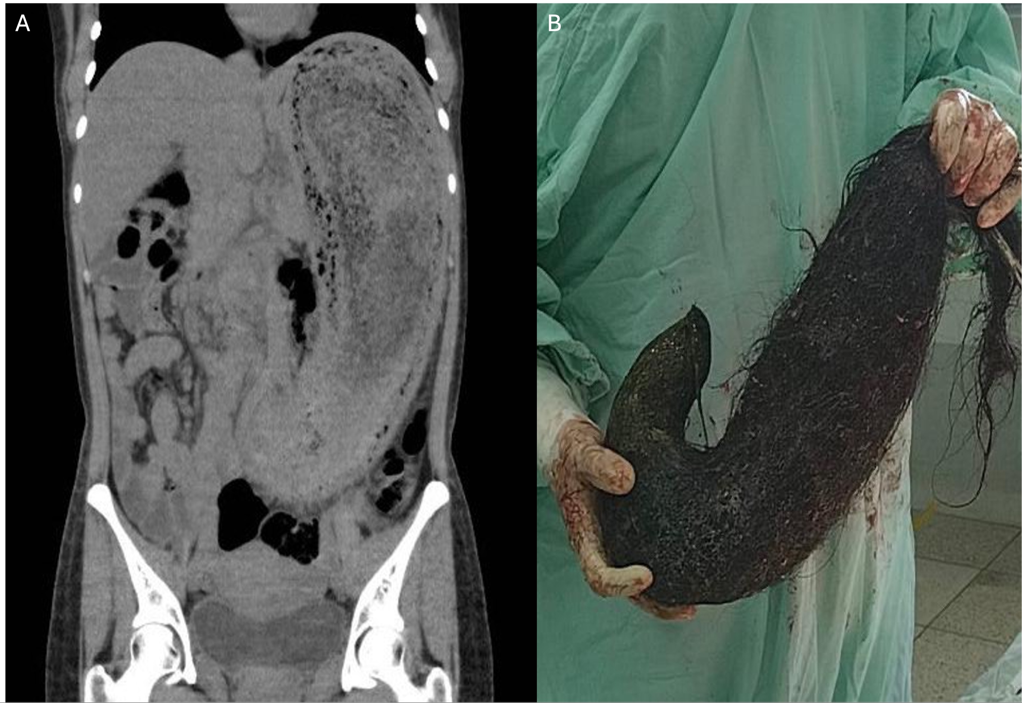
Case Description/Methods: A 24-year-old female patient with a history of trichophagia consulted the emergency department due to generalized abdominal pain associated with abdominal distention, nausea, vomiting, and absence of bowel movement in the last five days. On physical examination, she was found to have palpation of abdominal mass in the left upper and lower abdominal quadrants. Complete metabolic panel and complete blood count were within the normal range. Abdominal tomography revealed a stomach with a significant increase in size and occupied by hypodense contents with a swirling appearance (Figure 1A). Given the suspicion of obstruction of the gastrointestinal tract, it was decided to perform an open laparotomy with subsequent evidence of a large trichobezoar, which was removed without complications. (Figure 1B).
Discussion: Trichobezoars are congregations of compact masses composed of hair, which lacks sufficient friction surface for propulsion and is retained between the stomach folds. The presentation of gastric bezoar is insidious, nonspecific, and mainly asymptomatic. The most common clinical presentation is nausea, vomiting, epigastric pain, and weight loss. The most common finding in laboratories is evidence of anemia with vitamin B12 deficiency. Management can be endoscopic (biopsy and alligator forceps, lithotripters, needle cutter, snares of polypectomy, and lithotripsy) with or without fragmentation; however, most trichobezoars require surgical extraction (laparotomy with gastrostomy). This case could represent one of the most significant reported trichobezoar cases associated with gastric outlet syndrome.

Disclosures:
Juan Jose Chaves, MD1, Fernando Chamorro-Quiroz, MD2, Viviana Chaves-Cabezas, 3, José Chaves-Chamorro, MD2, María Camila Paredes, MD2. P3411 - Gastric Trichobezoar as a Presentation of Gastric Outlet Obstruction, ACG 2024 Annual Scientific Meeting Abstracts. Philadelphia, PA: American College of Gastroenterology.
1Norwalk Hospital, Norwalk, CT; 2Hospital Civil de Ipiales, Ipiales, Narino, Colombia; 3Fundacion Universitaria de Ciencias de la Salud, Bogota, Distrito Capital de Bogota, Colombia
Introduction: Bezoars are congregations or compact masses formed by matter accumulation, which results from the accumulation of material that is not digestible in the gastrointestinal tract, including hair, fiber-rich plant diet, and some pharmaceutical agents. They are relatively rare, with an incidence of less than 1% in the general population. Most cases occur in young women with a history of psychiatric pathology. Their location is mainly in the stomach; however, they can be located even in the small intestine.
Case Description/Methods: A 24-year-old female patient with a history of trichophagia consulted the emergency department due to generalized abdominal pain associated with abdominal distention, nausea, vomiting, and absence of bowel movement in the last five days. On physical examination, she was found to have palpation of abdominal mass in the left upper and lower abdominal quadrants. Complete metabolic panel and complete blood count were within the normal range. Abdominal tomography revealed a stomach with a significant increase in size and occupied by hypodense contents with a swirling appearance (Figure 1A). Given the suspicion of obstruction of the gastrointestinal tract, it was decided to perform an open laparotomy with subsequent evidence of a large trichobezoar, which was removed without complications. (Figure 1B).
Discussion: Trichobezoars are congregations of compact masses composed of hair, which lacks sufficient friction surface for propulsion and is retained between the stomach folds. The presentation of gastric bezoar is insidious, nonspecific, and mainly asymptomatic. The most common clinical presentation is nausea, vomiting, epigastric pain, and weight loss. The most common finding in laboratories is evidence of anemia with vitamin B12 deficiency. Management can be endoscopic (biopsy and alligator forceps, lithotripters, needle cutter, snares of polypectomy, and lithotripsy) with or without fragmentation; however, most trichobezoars require surgical extraction (laparotomy with gastrostomy). This case could represent one of the most significant reported trichobezoar cases associated with gastric outlet syndrome.

Figure: Figure 1. Gastric trichobezoar. An abdominal computed tomography (CT) scan revealed a significantly enlarged stomach occupied by hypodense contents, with a swirling appearance and thin walls (A). A surgical sample of a large trichobezoar measuring 20 cm x 20 cm and weighing 2,360 kg (B).
Disclosures:
Juan Jose Chaves indicated no relevant financial relationships.
Fernando Chamorro-Quiroz indicated no relevant financial relationships.
Viviana Chaves-Cabezas indicated no relevant financial relationships.
José Chaves-Chamorro indicated no relevant financial relationships.
María Camila Paredes indicated no relevant financial relationships.
Juan Jose Chaves, MD1, Fernando Chamorro-Quiroz, MD2, Viviana Chaves-Cabezas, 3, José Chaves-Chamorro, MD2, María Camila Paredes, MD2. P3411 - Gastric Trichobezoar as a Presentation of Gastric Outlet Obstruction, ACG 2024 Annual Scientific Meeting Abstracts. Philadelphia, PA: American College of Gastroenterology.
