Tuesday Poster Session
Category: Biliary/Pancreas
P3483 - Detection of Mural Nodules or Solid Components in Pancreatic Cystic Neoplasms: A Comparative Study of EUS vs CT/MRI
Tuesday, October 29, 2024
10:30 AM - 4:00 PM ET
Location: Exhibit Hall E

Has Audio

Ramez M. Ibrahim, MD
University of Texas MD Anderson Cancer Center
Houston, TX
Presenting Author(s)
Ramez M.. Ibrahim, MD, Suresh T.. Chari, MD, Matthew H. G.. Katz, MD, Michael P.. Kim, MD, Manoop S.. Bhutani, MD, FACG
University of Texas MD Anderson Cancer Center, Houston, TX
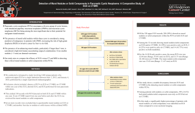
Introduction: Pancreatic cystic neoplasms (PCNs) encompass a diverse group of cystic lesions, with intraductal papillary mucinous neoplasms (IPMNs) and mucinous cystic neoplasms (MCNs) being among the most significant due to their potential for malignant transformation. The presence of mural/solid nodules within these cysts is considered a strong predictor of malignancy in patients with IPMN, increasing the risk of high-grade dysplasia (HGD) or invasive cancer by four- to six-fold. The presence of an enhancing mural nodule, particularly if larger than 5 mm, is considered a high-risk feature necessitating surgical consideration. Even smaller nodules (< 5 mm) are deemed worrisome. Our study aims to compare the efficacy of EUS versus CT and MRI in detecting these critical mural nodules or solid components within PCNs.
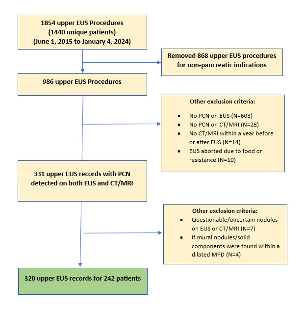
Methods: We conducted a retrospective study involving 1440 unique patients who underwent upper EUS at a single institution between June 1, 2015, and January 4, 2024, performed by a single experienced endosonographer. Exclusion criteria included: absence of PCN on EUS or CT/MRI, no CT/MRI within one year of the EUS, aborted EUS, and EUS performed for non-pancreatic indications. This yielded 330 records with PCN detected on both EUS and CT/MRI within one year. Every CT/MRI report from one year before and after the EUS was reviewed for mural nodules or solid components. Seven more records were excluded due to questionable mural nodules on EUS or CT/MRI, and another four due to nodules or solid masses within a dilated MPD.
Results: Of the 320 upper EUS records, 288 (90%) showed no mural nodules or solid components within the PCN on both EUS and CT/MRI. Among the 32 records showing mural nodules/solid components on EUS and/or CT/MRI, 16 (50%) were positive only on EUS, 2 (6.25%) were positive only on CT/MRI, and 14 (43.75%) were positive on both modalities. For the 16 EUS-only positive cases, the mean PCN size was 32.84 mm (Range: 9.4-91 mm) on EUS, and 33.75 mm (Range: 10-108 mm) on CT/MRI. The mean nodule/solid component size was 13.62 mm (Range: 3.1-67 mm) on EUS.
Discussion: Our study shows a notable discrepancy between EUS and CT/MRI in the detecting mural nodules or solid components within PCNs. Among patients with nodules or solid components, 50% (16/32) had mural nodules/solid components seen only on EUS but not on CT/MRI. In this study, a significantly higher percentage of patients with mural nodules or solid components were identified on EUS when compared to cross-sectional imaging.

Disclosures:
Ramez M.. Ibrahim, MD, Suresh T.. Chari, MD, Matthew H. G.. Katz, MD, Michael P.. Kim, MD, Manoop S.. Bhutani, MD, FACG. P3483 - Detection of Mural Nodules or Solid Components in Pancreatic Cystic Neoplasms: A Comparative Study of EUS vs CT/MRI, ACG 2024 Annual Scientific Meeting Abstracts. Philadelphia, PA: American College of Gastroenterology.
University of Texas MD Anderson Cancer Center, Houston, TX
Introduction: Pancreatic cystic neoplasms (PCNs) encompass a diverse group of cystic lesions, with intraductal papillary mucinous neoplasms (IPMNs) and mucinous cystic neoplasms (MCNs) being among the most significant due to their potential for malignant transformation. The presence of mural/solid nodules within these cysts is considered a strong predictor of malignancy in patients with IPMN, increasing the risk of high-grade dysplasia (HGD) or invasive cancer by four- to six-fold. The presence of an enhancing mural nodule, particularly if larger than 5 mm, is considered a high-risk feature necessitating surgical consideration. Even smaller nodules (< 5 mm) are deemed worrisome. Our study aims to compare the efficacy of EUS versus CT and MRI in detecting these critical mural nodules or solid components within PCNs.
Methods: We conducted a retrospective study involving 1440 unique patients who underwent upper EUS at a single institution between June 1, 2015, and January 4, 2024, performed by a single experienced endosonographer. Exclusion criteria included: absence of PCN on EUS or CT/MRI, no CT/MRI within one year of the EUS, aborted EUS, and EUS performed for non-pancreatic indications. This yielded 330 records with PCN detected on both EUS and CT/MRI within one year. Every CT/MRI report from one year before and after the EUS was reviewed for mural nodules or solid components. Seven more records were excluded due to questionable mural nodules on EUS or CT/MRI, and another four due to nodules or solid masses within a dilated MPD.
Results: Of the 320 upper EUS records, 288 (90%) showed no mural nodules or solid components within the PCN on both EUS and CT/MRI. Among the 32 records showing mural nodules/solid components on EUS and/or CT/MRI, 16 (50%) were positive only on EUS, 2 (6.25%) were positive only on CT/MRI, and 14 (43.75%) were positive on both modalities. For the 16 EUS-only positive cases, the mean PCN size was 32.84 mm (Range: 9.4-91 mm) on EUS, and 33.75 mm (Range: 10-108 mm) on CT/MRI. The mean nodule/solid component size was 13.62 mm (Range: 3.1-67 mm) on EUS.
Discussion: Our study shows a notable discrepancy between EUS and CT/MRI in the detecting mural nodules or solid components within PCNs. Among patients with nodules or solid components, 50% (16/32) had mural nodules/solid components seen only on EUS but not on CT/MRI. In this study, a significantly higher percentage of patients with mural nodules or solid components were identified on EUS when compared to cross-sectional imaging.

Figure: Study Flowchart
Disclosures:
Ramez Ibrahim indicated no relevant financial relationships.
Suresh Chari indicated no relevant financial relationships.
Matthew Katz indicated no relevant financial relationships.
Michael Kim indicated no relevant financial relationships.
Manoop Bhutani indicated no relevant financial relationships.
Ramez M.. Ibrahim, MD, Suresh T.. Chari, MD, Matthew H. G.. Katz, MD, Michael P.. Kim, MD, Manoop S.. Bhutani, MD, FACG. P3483 - Detection of Mural Nodules or Solid Components in Pancreatic Cystic Neoplasms: A Comparative Study of EUS vs CT/MRI, ACG 2024 Annual Scientific Meeting Abstracts. Philadelphia, PA: American College of Gastroenterology.
