Tuesday Poster Session
Category: Biliary/Pancreas
P3549 - A Case Report of Acute Pancreatitis in Food-Induced Anaphylaxis
Tuesday, October 29, 2024
10:30 AM - 4:00 PM ET
Location: Exhibit Hall E

Has Audio
- JW
Jennifer Wiese, MD
Joan C. Edwards School of Medicine, Marshall University
Huntington, WV
Presenting Author(s)
Jennifer Wiese, MD1, Bassel Dakkak, MD2, Onyinye Ugonabo, MD3, Mohammed El-Dallal, MD, MMSc1, Wesam M. Frandah, MD2
1Joan C. Edwards School of Medicine, Marshall University, Huntington, WV; 2Marshall University Joan C. Edwards School of Medicine, Huntington, WV; 3Marshall University, Huntington, WV
Introduction: Acute pancreatitis (AP) is estimated to be 300,000 emergency department visits each year and is one of the leading causes of gastrointestinal-related hospital admissions. Most common causes of pancreatitis include toxic, metabolic, or mechanical etiologies. Very few cases of acute pancreatitis have been linked to food-induced anaphylaxis. Our case will enrich the literature on this matter and will highlight how food allergy-induced pancreatitis should be considered in idiopathic pancreatitis cases, which could lead to more targeted diagnoses and improved management strategies.
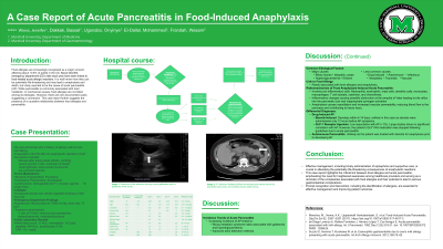
Case Description/Methods: A 54-year-old female with a history of asthma and hypothyroidism, known allergies to Percocet, latex, milk, azithromycin, and contrast, presented with anaphylactic reaction to food. She reported feeling nauseous after eating steak alfredo followed by vomiting, severe pruritus, hives, shortness of breath, chest tightness, and loose bowel movement along with a pre-syncopal episode. Initial blood pressure was 75/66 mmHg and heart rate was 75 bpm. She was treated with epinephrine, solumedrol and intravenous fluid (IV). Her vitals improved, and she was continued with IV fluid and was admitted for further monitoring. Initial laboratory findings were grossly unremarkable. Twelve hours later, she started complaining of abdominal pain. Abdominal Ultrasound was unremarkable and negative for cholelithiasis. CT abdomen displayed diffuse fat stranding and edema across the pancreatic body, head, and uncinate process,
concerning for acute pancreatitis. Repeat labs showed elevated lipase and amylase, consistent with acute pancreatitis. The patient was managed supportively for acute pancreatitis with IV fluid at 1.5 ml/kg/h for 24 hours and analgesic. Her abdominal pain improved within 72 hours, and she began to tolerate diet. She was discharged with follow-up appointments.
Discussion: This case highlights the significance of diagnosing AP in patients with anaphylaxis. Outcomes from acute pancreatitis are influenced by risk stratification, fluid, and nutritional management, and follow-up care and risk reduction strategies. Prompt diagnosis, early recognition of food allergy- induced acute pancreatitis and stratification of severity with proper management can significantly reduce morbidity and mortality.

Note: The table for this abstract can be viewed in the ePoster Gallery section of the ACG 2024 ePoster Site or in The American Journal of Gastroenterology's abstract supplement issue, both of which will be available starting October 27, 2024.
Disclosures:
Jennifer Wiese, MD1, Bassel Dakkak, MD2, Onyinye Ugonabo, MD3, Mohammed El-Dallal, MD, MMSc1, Wesam M. Frandah, MD2. P3549 - A Case Report of Acute Pancreatitis in Food-Induced Anaphylaxis, ACG 2024 Annual Scientific Meeting Abstracts. Philadelphia, PA: American College of Gastroenterology.
1Joan C. Edwards School of Medicine, Marshall University, Huntington, WV; 2Marshall University Joan C. Edwards School of Medicine, Huntington, WV; 3Marshall University, Huntington, WV
Introduction: Acute pancreatitis (AP) is estimated to be 300,000 emergency department visits each year and is one of the leading causes of gastrointestinal-related hospital admissions. Most common causes of pancreatitis include toxic, metabolic, or mechanical etiologies. Very few cases of acute pancreatitis have been linked to food-induced anaphylaxis. Our case will enrich the literature on this matter and will highlight how food allergy-induced pancreatitis should be considered in idiopathic pancreatitis cases, which could lead to more targeted diagnoses and improved management strategies.
Case Description/Methods: A 54-year-old female with a history of asthma and hypothyroidism, known allergies to Percocet, latex, milk, azithromycin, and contrast, presented with anaphylactic reaction to food. She reported feeling nauseous after eating steak alfredo followed by vomiting, severe pruritus, hives, shortness of breath, chest tightness, and loose bowel movement along with a pre-syncopal episode. Initial blood pressure was 75/66 mmHg and heart rate was 75 bpm. She was treated with epinephrine, solumedrol and intravenous fluid (IV). Her vitals improved, and she was continued with IV fluid and was admitted for further monitoring. Initial laboratory findings were grossly unremarkable. Twelve hours later, she started complaining of abdominal pain. Abdominal Ultrasound was unremarkable and negative for cholelithiasis. CT abdomen displayed diffuse fat stranding and edema across the pancreatic body, head, and uncinate process,
concerning for acute pancreatitis. Repeat labs showed elevated lipase and amylase, consistent with acute pancreatitis. The patient was managed supportively for acute pancreatitis with IV fluid at 1.5 ml/kg/h for 24 hours and analgesic. Her abdominal pain improved within 72 hours, and she began to tolerate diet. She was discharged with follow-up appointments.
Discussion: This case highlights the significance of diagnosing AP in patients with anaphylaxis. Outcomes from acute pancreatitis are influenced by risk stratification, fluid, and nutritional management, and follow-up care and risk reduction strategies. Prompt diagnosis, early recognition of food allergy- induced acute pancreatitis and stratification of severity with proper management can significantly reduce morbidity and mortality.

Figure: Image 1: Ultrasound the abdomen showing normal gallbladder with no gallstones (white arrow).
CT abdomen displaying diffuse fat stranding and edema around the pancreatic body, head, and uncinate process (white arrow).
CT abdomen displaying diffuse fat stranding and edema around the pancreatic body, head, and uncinate process (white arrow).
Note: The table for this abstract can be viewed in the ePoster Gallery section of the ACG 2024 ePoster Site or in The American Journal of Gastroenterology's abstract supplement issue, both of which will be available starting October 27, 2024.
Disclosures:
Jennifer Wiese indicated no relevant financial relationships.
Bassel Dakkak indicated no relevant financial relationships.
Onyinye Ugonabo indicated no relevant financial relationships.
Mohammed El-Dallal indicated no relevant financial relationships.
Wesam Frandah: Endogastric solution – Consultant.
Jennifer Wiese, MD1, Bassel Dakkak, MD2, Onyinye Ugonabo, MD3, Mohammed El-Dallal, MD, MMSc1, Wesam M. Frandah, MD2. P3549 - A Case Report of Acute Pancreatitis in Food-Induced Anaphylaxis, ACG 2024 Annual Scientific Meeting Abstracts. Philadelphia, PA: American College of Gastroenterology.
