Tuesday Poster Session
Category: Biliary/Pancreas
P3564 - Pancreatitis and Obstructive Jaundice as Presenting Symptoms for Ampullary Pancreatic Neuroendocrine Tumor
Tuesday, October 29, 2024
10:30 AM - 4:00 PM ET
Location: Exhibit Hall E

Has Audio
- VM
Viraj Modi, DO
HCA Florida Healthcare
riverview, FL
Presenting Author(s)
Viraj D. Modi, DO1, Muhammad Qasim Ali, MD2, Christina Maldonado, MD2, Marwa Hegagi, MD2
1HCA Florida Healthcare, Riverview, FL; 2HCA Florida Healthcare, Brandon, FL
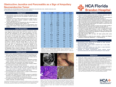
Introduction: Neuroendocrine tumors (NETs) are rare and can be classified as either functional or nonfunctional. Functional NETs present with various hormonal secretions and associated clinical syndromes. In contrast, nonfunctional NETs primarily present due to mass effect, often at a later stage. Here, we present a rare case of a nonfunctional ampullary NET manifesting as pancreatitis.
Case Description/Methods: A 61-year-old male with hypertension presented to the ED with severe, sharp epigastric pain rated as a 10/10 radiating to his back for 24 hours. The pain worsened with meals and was accompanied by nausea, vomiting, constipation, and jaundice. Self-medication with PPIs provided no relief. The patient denied alcohol use or a history of gallstones. On examination, he had tenderness in the epigastric and right upper quadrant without rebound. Lab results showed total bilirubin 5.9, AST 137, ALT 326, ALP 339, and Lipase 622. A CT scan indicated pancreatitis with peripancreatic lymph node enlargement and an 8 mm biliary tree dilatation without gallstones. An MRCP showed mild soft tissue prominence at the pancreatic head and a mildly distended gallbladder without filling defects for which an ERCP identified a large fungating mass at the ampulla of Vater. Due to abnormal anatomy, cannulation was aborted. Biopsies confirmed high-grade neuroendocrine carcinoma with high Ki proliferative index. Percutaneous transhepatic biliary drainage and catheter placement were performed. The patient was scheduled for a pancreaticoduodenectomy, which was aborted as the tumor was found to be encasing the SMA/SMV. The patient was to undergo adjuvant chemotherapy with cisplatin, etoposide, and radiation therapy.
Discussion: Nonfunctioning neuroendocrine tumors often present later with symptoms related to local compression or metastasis, such as abdominal pain and weight loss, and less frequently with obstructive jaundice. In our case, the patient presented with pancreatitis due to mass effect at the ampulla, leading to poor pancreatic enzyme release into the duodenal lumen. A biopsy confirmed high-grade neuroendocrine carcinoma characterized by small to medium-sized nuclei, scant cytoplasm, positive CD56 and synaptophysin staining, and a high Ki-67 index of 100%. Given the bulkiness of the tumor, which precluded resection, adjuvant chemotherapy was deemed the most appropriate treatment.

Note: The table for this abstract can be viewed in the ePoster Gallery section of the ACG 2024 ePoster Site or in The American Journal of Gastroenterology's abstract supplement issue, both of which will be available starting October 27, 2024.
Disclosures:
Viraj D. Modi, DO1, Muhammad Qasim Ali, MD2, Christina Maldonado, MD2, Marwa Hegagi, MD2. P3564 - Pancreatitis and Obstructive Jaundice as Presenting Symptoms for Ampullary Pancreatic Neuroendocrine Tumor, ACG 2024 Annual Scientific Meeting Abstracts. Philadelphia, PA: American College of Gastroenterology.
1HCA Florida Healthcare, Riverview, FL; 2HCA Florida Healthcare, Brandon, FL
Introduction: Neuroendocrine tumors (NETs) are rare and can be classified as either functional or nonfunctional. Functional NETs present with various hormonal secretions and associated clinical syndromes. In contrast, nonfunctional NETs primarily present due to mass effect, often at a later stage. Here, we present a rare case of a nonfunctional ampullary NET manifesting as pancreatitis.
Case Description/Methods: A 61-year-old male with hypertension presented to the ED with severe, sharp epigastric pain rated as a 10/10 radiating to his back for 24 hours. The pain worsened with meals and was accompanied by nausea, vomiting, constipation, and jaundice. Self-medication with PPIs provided no relief. The patient denied alcohol use or a history of gallstones. On examination, he had tenderness in the epigastric and right upper quadrant without rebound. Lab results showed total bilirubin 5.9, AST 137, ALT 326, ALP 339, and Lipase 622. A CT scan indicated pancreatitis with peripancreatic lymph node enlargement and an 8 mm biliary tree dilatation without gallstones. An MRCP showed mild soft tissue prominence at the pancreatic head and a mildly distended gallbladder without filling defects for which an ERCP identified a large fungating mass at the ampulla of Vater. Due to abnormal anatomy, cannulation was aborted. Biopsies confirmed high-grade neuroendocrine carcinoma with high Ki proliferative index. Percutaneous transhepatic biliary drainage and catheter placement were performed. The patient was scheduled for a pancreaticoduodenectomy, which was aborted as the tumor was found to be encasing the SMA/SMV. The patient was to undergo adjuvant chemotherapy with cisplatin, etoposide, and radiation therapy.
Discussion: Nonfunctioning neuroendocrine tumors often present later with symptoms related to local compression or metastasis, such as abdominal pain and weight loss, and less frequently with obstructive jaundice. In our case, the patient presented with pancreatitis due to mass effect at the ampulla, leading to poor pancreatic enzyme release into the duodenal lumen. A biopsy confirmed high-grade neuroendocrine carcinoma characterized by small to medium-sized nuclei, scant cytoplasm, positive CD56 and synaptophysin staining, and a high Ki-67 index of 100%. Given the bulkiness of the tumor, which precluded resection, adjuvant chemotherapy was deemed the most appropriate treatment.

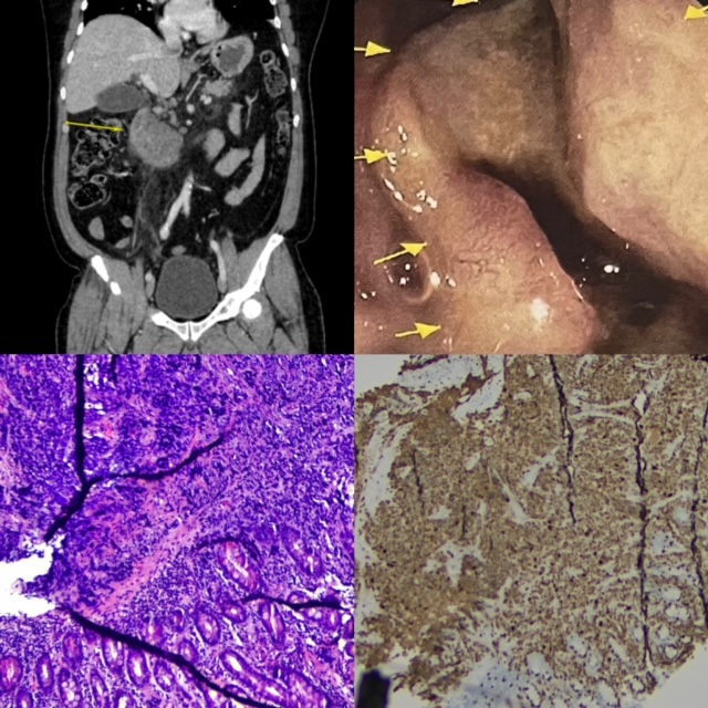
Figure: Top Left: CT Abdomen/Pelvis taken during admission, with heterogenous mass near head of pancreas. Top Right: Gross image of NET at ampulla of Vater during EGD/ERCP. Bottom Left: H&E stain at 10x magnitude showing infiltrative tumor of small cells with scant cytoplasm, numerous mitotic figures and scattered necrosis. Bottom Right: Ki67 stain demonstrating staining in 100% of tumor cells consistent with high-grade dysplasia.
Note: The table for this abstract can be viewed in the ePoster Gallery section of the ACG 2024 ePoster Site or in The American Journal of Gastroenterology's abstract supplement issue, both of which will be available starting October 27, 2024.
Disclosures:
Viraj Modi indicated no relevant financial relationships.
Muhammad Qasim Ali indicated no relevant financial relationships.
Christina Maldonado indicated no relevant financial relationships.
Marwa Hegagi indicated no relevant financial relationships.
Viraj D. Modi, DO1, Muhammad Qasim Ali, MD2, Christina Maldonado, MD2, Marwa Hegagi, MD2. P3564 - Pancreatitis and Obstructive Jaundice as Presenting Symptoms for Ampullary Pancreatic Neuroendocrine Tumor, ACG 2024 Annual Scientific Meeting Abstracts. Philadelphia, PA: American College of Gastroenterology.
