Tuesday Poster Session
Category: Biliary/Pancreas
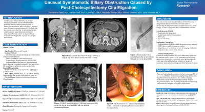
P3567 - Unusual Symptomatic Biliary Obstruction Caused by Post-Cholecystectomy Clip Migration
Tuesday, October 29, 2024
10:30 AM - 4:00 PM ET
Location: Exhibit Hall E

Has Audio
- SP
Sarvanand Patel, MD
Kaiser Permanente
Los Angeles, CA
Presenting Author(s)
Sarvanand Patel, MD, Steven Park, MD, Cynthia Liu, MD, Harpreet Sekhon, MD, Manav Sharma, MD, John Iskander, MD
Kaiser Permanente, Los Angeles, CA
Introduction: Laparoscopic cholecystectomy (LC) is the minimally invasive standard treatment for gallstone disease associated with reduced post-operative pain and shorter hospital stays. Complications of LC include bile duct injury, infection, and rare yet significant post-cholecystectomy clip migration (PCCM) that requires endoscopic intervention.
Case Description/Methods: A 54-year-old woman presented with 2 days of post-prandial right upper-quadrant pain. She had a history of cholelithiasis, acute cholecystitis, and choledocholithiasis treated with ERCP and LC in 2006. The operative report noted a severely inflamed gallbladder and dense adhesions that made isolating the cystic duct challenging. Four clips were applied to the cystic duct, three clips were applied to the cystic artery, and additional multiple clips (unspecified) placed in the surrounding adhesions. Also, two years following the surgery, the patient developed an intra-abdominal abscess and a right flank fluid collection evacuated in the OR with findings of 10 gallstones and a surgical clip.
At the current presentation, our patient was afebrile (36.6 °C) with a blood pressure 100/69mmHg, pulse of 82bpm, respiratory rate at 18/min. Her physical exam was remarkable for epigastric pain, though Murphy’s sign was negative.
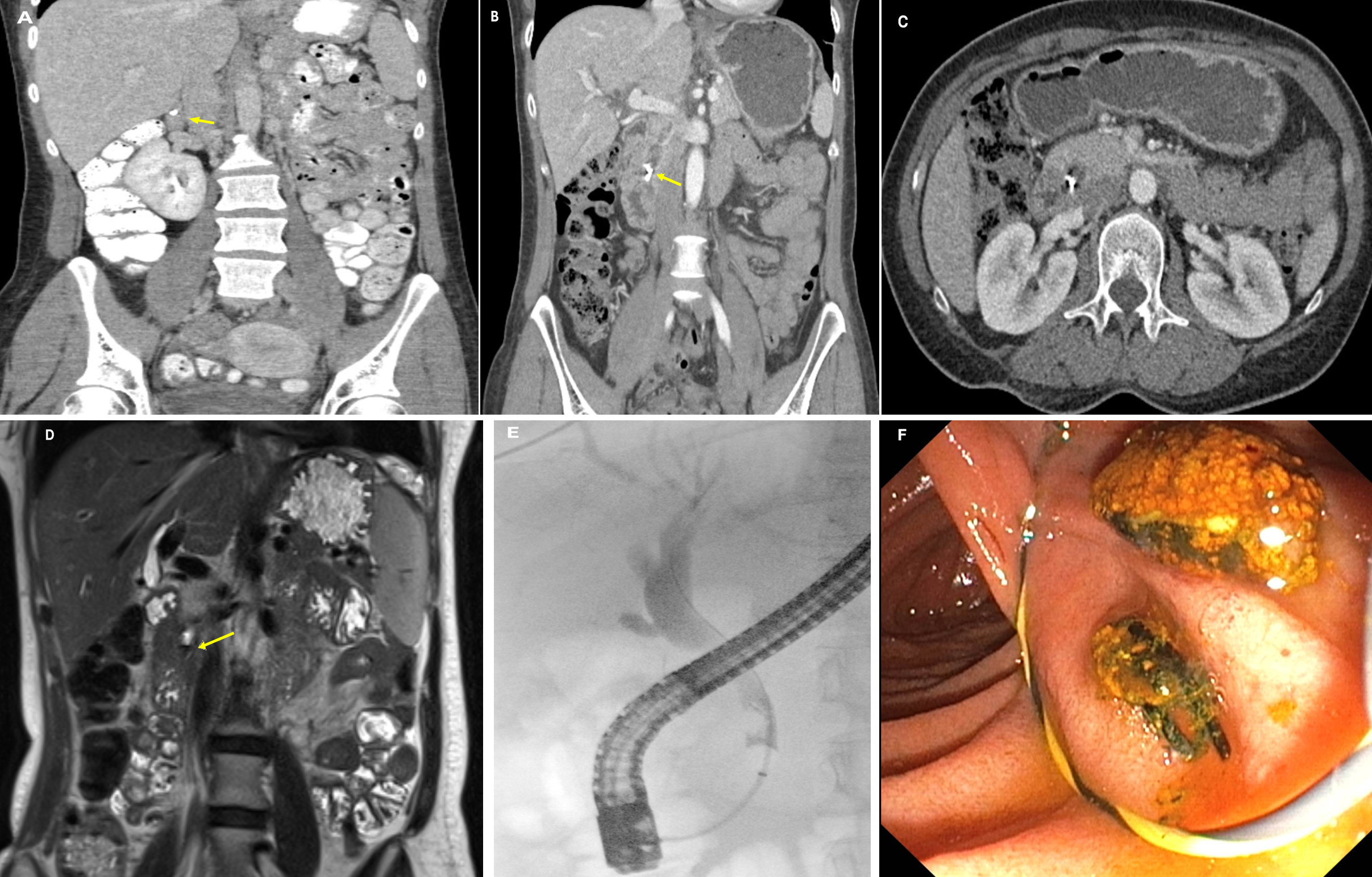
Lab results showed a white count of 5.7 [4-11x1000/mcL], alanine transaminase of 1100 [< =54U/L], aspartate transaminase of 861 [< =30U/L], alkaline phosphatase of 228 [< =125U/L], total bilirubin of 2.0 [< =0.2mg/dL], and normal lipase. Abdominal CT with IV contrast showed surgical clips adjacent to a dilated common bile duct of 10mm compared to the previous CT (Figure A, B, C). The pancreas was unremarkable. MRCP showed metallic artifacts adjacent to the distal CBD (Figure D).
Patient underwent ERCP for further evaluation at which time cholangiogram revealed a clip and filling defects in the bile duct (Figure E). ERCP with sphincteroplasty and balloon sweep was used to extract the cholecystectomy clip (Figure F).
Discussion: Our case demonstrates a symptomatic biliary obstruction from post-cholecystectomy clip migration 16 years after a challenging LC. The reported median time of clip migration is 2 years, ranging from 11 days to 20 years after cholecystectomy. Close inspection of the imaging studies can distinguish between primary CBD stones from clip migration-related complications. Inaccurate clip placements, local suppurative inflammation, bile leaks, and local infections are noted risk factors.

Disclosures:
Sarvanand Patel, MD, Steven Park, MD, Cynthia Liu, MD, Harpreet Sekhon, MD, Manav Sharma, MD, John Iskander, MD. P3567 - Unusual Symptomatic Biliary Obstruction Caused by Post-Cholecystectomy Clip Migration, ACG 2024 Annual Scientific Meeting Abstracts. Philadelphia, PA: American College of Gastroenterology.
Kaiser Permanente, Los Angeles, CA
Introduction: Laparoscopic cholecystectomy (LC) is the minimally invasive standard treatment for gallstone disease associated with reduced post-operative pain and shorter hospital stays. Complications of LC include bile duct injury, infection, and rare yet significant post-cholecystectomy clip migration (PCCM) that requires endoscopic intervention.
Case Description/Methods: A 54-year-old woman presented with 2 days of post-prandial right upper-quadrant pain. She had a history of cholelithiasis, acute cholecystitis, and choledocholithiasis treated with ERCP and LC in 2006. The operative report noted a severely inflamed gallbladder and dense adhesions that made isolating the cystic duct challenging. Four clips were applied to the cystic duct, three clips were applied to the cystic artery, and additional multiple clips (unspecified) placed in the surrounding adhesions. Also, two years following the surgery, the patient developed an intra-abdominal abscess and a right flank fluid collection evacuated in the OR with findings of 10 gallstones and a surgical clip.
At the current presentation, our patient was afebrile (36.6 °C) with a blood pressure 100/69mmHg, pulse of 82bpm, respiratory rate at 18/min. Her physical exam was remarkable for epigastric pain, though Murphy’s sign was negative.
Lab results showed a white count of 5.7 [4-11x1000/mcL], alanine transaminase of 1100 [< =54U/L], aspartate transaminase of 861 [< =30U/L], alkaline phosphatase of 228 [< =125U/L], total bilirubin of 2.0 [< =0.2mg/dL], and normal lipase. Abdominal CT with IV contrast showed surgical clips adjacent to a dilated common bile duct of 10mm compared to the previous CT (Figure A, B, C). The pancreas was unremarkable. MRCP showed metallic artifacts adjacent to the distal CBD (Figure D).
Patient underwent ERCP for further evaluation at which time cholangiogram revealed a clip and filling defects in the bile duct (Figure E). ERCP with sphincteroplasty and balloon sweep was used to extract the cholecystectomy clip (Figure F).
Discussion: Our case demonstrates a symptomatic biliary obstruction from post-cholecystectomy clip migration 16 years after a challenging LC. The reported median time of clip migration is 2 years, ranging from 11 days to 20 years after cholecystectomy. Close inspection of the imaging studies can distinguish between primary CBD stones from clip migration-related complications. Inaccurate clip placements, local suppurative inflammation, bile leaks, and local infections are noted risk factors.

Figure: Figure A: Prior coronal CT with contrast showing initial surgical clip location.
Figure B & C: Coronal and Axial CT images showing a surgical clip in the distal common bile duct (arrow)
Figure D: MRCP shows dilatation of CBD and intrahepatic bile ducts with clip in the distal CBD. with no definite choledocholithiasis
Figure E: Fluoroscopy with a metallic hue within an amorphous filling defect in the distal CBD
Figure F: ERCP extraction of a surgical clip embedded within a gallstone. A separate gallstone is noted above the surgical clip.
Disclosures:
Sarvanand Patel indicated no relevant financial relationships.
Steven Park indicated no relevant financial relationships.
Cynthia Liu indicated no relevant financial relationships.
Harpreet Sekhon indicated no relevant financial relationships.
Manav Sharma indicated no relevant financial relationships.
John Iskander indicated no relevant financial relationships.
Sarvanand Patel, MD, Steven Park, MD, Cynthia Liu, MD, Harpreet Sekhon, MD, Manav Sharma, MD, John Iskander, MD. P3567 - Unusual Symptomatic Biliary Obstruction Caused by Post-Cholecystectomy Clip Migration, ACG 2024 Annual Scientific Meeting Abstracts. Philadelphia, PA: American College of Gastroenterology.
