Tuesday Poster Session
Category: Biliary/Pancreas
P3574 - Unmasking The Pancreatic Phantom
Tuesday, October 29, 2024
10:30 AM - 4:00 PM ET
Location: Exhibit Hall E

Has Audio

Geraldine N. Nabeta, MS MD
University of Connecticut Health Center
Farmington, CT
Presenting Author(s)
Geraldine N. Nabeta, MS MD1, Amanda Frondella, MD2, Ramachandra Illindala, MD3
1University of Connecticut Health Center, Farmington, CT; 2UConn John Dempsey Hospital, Farmington, CT; 3Hospital of Central Connecticut, New Britain, CT
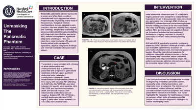
Introduction: Metastatic pancreatic cancer represents a severe and often fatal condition, characterized by its aggressive nature. Predominantly originating in the head of the pancreas, its typical clinical manifestations include epigastric pain, jaundice, diarrhea, steatorrhea, nausea, and vomiting. Despite the availability of advanced abdominal imaging modalities with diagnostic sensitivities averaging 90%, certain cases present significant diagnostic challenges. Here, we describe a case involving a 59-year-old female patient with nonspecific symptoms, atypical diagnostic findings, and minimal risk factors for pancreatic cancer.
Case Description/Methods: The patient, a never-smoker with a history of acute cholecystitis and cholecystectomy due to gallstones, presented with progressive generalized weakness and right upper quadrant abdominal pain. Laboratory investigations revealed a mixed hepatocellular and cholestatic pattern of liver injury, with elevated AST (368 U/L), ALT (427 U/L), ALP (371 U/L), and total bilirubin (2.2 mg/dL). Viral hepatitis, HSV, CMV, EBV, and autoimmune markers (ANA, anti-smooth muscle, anti-mitochondrial antibodies) were all negative. Notably, CA 19-9 (472 U/mL), and IgG levels were elevated (1726 mg/dL), while other tumor markers (CA 125, CEA) were unremarkable.
Initial abdominal US and CT scans were unremarkable except for a subcentimeter hypodense lesion in the pancreatic body, described as a pancreatic cyst or benign IPMN. Follow-up MRI/MRCP indicated several rim-enhancing hepatic lesions initially presumed to be microabscesses. The initial liver biopsy suggested a cholangiopathy pattern.
Subsequent biopsies revealed a preliminary suspicion for pancreatic adenocarcinoma with benign liver lesions. Given the high clinical suspicion for malignancy, a further liver biopsy was conducted, which confirmed adenocarcinoma of pancreatobiliary origin. The patient commenced palliative chemotherapy, given the incurable nature of her cancer.
Discussion: This case underscores the complexities involved in diagnosing metastatic pancreatic cancer, especially when initial presentations and diagnostic tests are atypical. The importance of re-evaluation, regular follow-up, and the correlation between clinical judgment and patient presentation cannot be overstated. This report highlights the necessity for healthcare providers to maintain a high index of suspicion and to pursue comprehensive diagnostic strategies in similar challenging cases
Disclosures:
Geraldine N. Nabeta, MS MD1, Amanda Frondella, MD2, Ramachandra Illindala, MD3. P3574 - Unmasking The Pancreatic Phantom, ACG 2024 Annual Scientific Meeting Abstracts. Philadelphia, PA: American College of Gastroenterology.
1University of Connecticut Health Center, Farmington, CT; 2UConn John Dempsey Hospital, Farmington, CT; 3Hospital of Central Connecticut, New Britain, CT
Introduction: Metastatic pancreatic cancer represents a severe and often fatal condition, characterized by its aggressive nature. Predominantly originating in the head of the pancreas, its typical clinical manifestations include epigastric pain, jaundice, diarrhea, steatorrhea, nausea, and vomiting. Despite the availability of advanced abdominal imaging modalities with diagnostic sensitivities averaging 90%, certain cases present significant diagnostic challenges. Here, we describe a case involving a 59-year-old female patient with nonspecific symptoms, atypical diagnostic findings, and minimal risk factors for pancreatic cancer.
Case Description/Methods: The patient, a never-smoker with a history of acute cholecystitis and cholecystectomy due to gallstones, presented with progressive generalized weakness and right upper quadrant abdominal pain. Laboratory investigations revealed a mixed hepatocellular and cholestatic pattern of liver injury, with elevated AST (368 U/L), ALT (427 U/L), ALP (371 U/L), and total bilirubin (2.2 mg/dL). Viral hepatitis, HSV, CMV, EBV, and autoimmune markers (ANA, anti-smooth muscle, anti-mitochondrial antibodies) were all negative. Notably, CA 19-9 (472 U/mL), and IgG levels were elevated (1726 mg/dL), while other tumor markers (CA 125, CEA) were unremarkable.
Initial abdominal US and CT scans were unremarkable except for a subcentimeter hypodense lesion in the pancreatic body, described as a pancreatic cyst or benign IPMN. Follow-up MRI/MRCP indicated several rim-enhancing hepatic lesions initially presumed to be microabscesses. The initial liver biopsy suggested a cholangiopathy pattern.
Subsequent biopsies revealed a preliminary suspicion for pancreatic adenocarcinoma with benign liver lesions. Given the high clinical suspicion for malignancy, a further liver biopsy was conducted, which confirmed adenocarcinoma of pancreatobiliary origin. The patient commenced palliative chemotherapy, given the incurable nature of her cancer.
Discussion: This case underscores the complexities involved in diagnosing metastatic pancreatic cancer, especially when initial presentations and diagnostic tests are atypical. The importance of re-evaluation, regular follow-up, and the correlation between clinical judgment and patient presentation cannot be overstated. This report highlights the necessity for healthcare providers to maintain a high index of suspicion and to pursue comprehensive diagnostic strategies in similar challenging cases
Disclosures:
Geraldine Nabeta indicated no relevant financial relationships.
Amanda Frondella indicated no relevant financial relationships.
Ramachandra Illindala indicated no relevant financial relationships.
Geraldine N. Nabeta, MS MD1, Amanda Frondella, MD2, Ramachandra Illindala, MD3. P3574 - Unmasking The Pancreatic Phantom, ACG 2024 Annual Scientific Meeting Abstracts. Philadelphia, PA: American College of Gastroenterology.
