Tuesday Poster Session
Category: Biliary/Pancreas
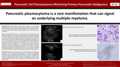
P3575 - Pancreatic Tail Plasmacytoma Mimicking Primary Pancreatic Malignancy
Tuesday, October 29, 2024
10:30 AM - 4:00 PM ET
Location: Exhibit Hall E

Has Audio

David Weinstein, MD
University of Chicago, NorthShore Internal Medicine
Evanston, IL
Presenting Author(s)
David Weinstein, MD1, Sarah Burroughs, DO2, Robert Toelke, MD3, Raza Muhammad, DO4, Farhan Quader, MD3
1University of Chicago, NorthShore Internal Medicine, Evanston, IL; 2University of Chicago, Northshore University Healthsystem, Evanston, IL; 3NorthShore University HealthSystem, Evanston, IL; 4University of Chicago Northshore, Evanston, IL
Introduction: Extramedullary plasmacytomas (EMPs) are hematological malignancies that occur in mucosal surfaces outside the bone marrow. They are rare, with about 10% occurring in the gastrointestinal tract. Presented here is a rare case of pancreatic tail EMP initially thought to be a primary pancreatic malignancy.
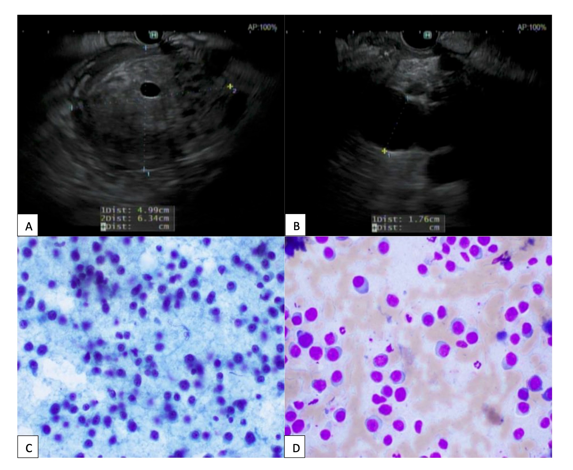
Case Description/Methods: An 87-year-old male with a past medical history of multiple myeloma in remission presented with 7 months of diarrhea. During this time, he also reported a 20-pound weight loss and progressive, severe back pain. Laboratory workup revealed white blood cell count of 7.8 x 10³/µL, hemoglobin of 14.5 g/dL, and platelets of 157 x 103/µL. Blood glucose testing showed elevations up to 197 mg/dL with an A1c of 6.7%. Stool testing revealed low fecal elastase levels, which raised concern for pancreatic insufficiency. He subsequently underwent CT of the abdomen with contrast which revealed multiple high-density masses in the pleura, pancreas, and right sciatic region. MRI/MRCP confirmed an 8 cm x 6 cm pancreatic tail mass along with numerous cysts involving the pancreatic tail and head, and diffuse pancreatic duct dilatation measuring up to 1.7 cm. CA 19-9 was normal (< 2.0 U/mL). EUS was performed which revealed an irregular mass within the pancreatic tail measuring 6 cm x 5.5 cm along with a dilated pancreatic duct measuring 1.7 cm in diameter. Initial biopsies taken were inconclusive but not suggestive of adenocarcinoma. Repeat endoscopic ultrasound revealed the same pancreatic tail mass measuring 7.1 cm x 6.2 cm along with pancreatic duct dilatation measuring 1 cm in diameter. Biopsies confirmed plasmacytoma of the pancreatic tail. He was then started on radiation with follow up PET/CT scan demonstrating significant regression of both masses.
Discussion: EMPs are rare, accounting for about 4% of all plasma cell neoplasms, and only a few cases demonstrate pancreatic tail involvement. Pancreatic plasmacytomas are typically seen in patients with concurrent, or preceding, MM, and 30% of patients develop MM within 10 years of diagnosis. Our patient had poor prognostic factors, including a tumor size > 4 cm in diameter, age of onset > 50 years, and EMP occurring outside the head and neck. The differential included plasmacytoma, lymphoma, amyloidosis, adenocarcinoma, and neuroendocrine tumor. Treatment for pancreatic plasmacytoma involves radiation, chemotherapy, and/or surgical resection based on its location, but there appears to be no standardized approach given its rarity.

Disclosures:
David Weinstein, MD1, Sarah Burroughs, DO2, Robert Toelke, MD3, Raza Muhammad, DO4, Farhan Quader, MD3. P3575 - Pancreatic Tail Plasmacytoma Mimicking Primary Pancreatic Malignancy, ACG 2024 Annual Scientific Meeting Abstracts. Philadelphia, PA: American College of Gastroenterology.
1University of Chicago, NorthShore Internal Medicine, Evanston, IL; 2University of Chicago, Northshore University Healthsystem, Evanston, IL; 3NorthShore University HealthSystem, Evanston, IL; 4University of Chicago Northshore, Evanston, IL
Introduction: Extramedullary plasmacytomas (EMPs) are hematological malignancies that occur in mucosal surfaces outside the bone marrow. They are rare, with about 10% occurring in the gastrointestinal tract. Presented here is a rare case of pancreatic tail EMP initially thought to be a primary pancreatic malignancy.
Case Description/Methods: An 87-year-old male with a past medical history of multiple myeloma in remission presented with 7 months of diarrhea. During this time, he also reported a 20-pound weight loss and progressive, severe back pain. Laboratory workup revealed white blood cell count of 7.8 x 10³/µL, hemoglobin of 14.5 g/dL, and platelets of 157 x 103/µL. Blood glucose testing showed elevations up to 197 mg/dL with an A1c of 6.7%. Stool testing revealed low fecal elastase levels, which raised concern for pancreatic insufficiency. He subsequently underwent CT of the abdomen with contrast which revealed multiple high-density masses in the pleura, pancreas, and right sciatic region. MRI/MRCP confirmed an 8 cm x 6 cm pancreatic tail mass along with numerous cysts involving the pancreatic tail and head, and diffuse pancreatic duct dilatation measuring up to 1.7 cm. CA 19-9 was normal (< 2.0 U/mL). EUS was performed which revealed an irregular mass within the pancreatic tail measuring 6 cm x 5.5 cm along with a dilated pancreatic duct measuring 1.7 cm in diameter. Initial biopsies taken were inconclusive but not suggestive of adenocarcinoma. Repeat endoscopic ultrasound revealed the same pancreatic tail mass measuring 7.1 cm x 6.2 cm along with pancreatic duct dilatation measuring 1 cm in diameter. Biopsies confirmed plasmacytoma of the pancreatic tail. He was then started on radiation with follow up PET/CT scan demonstrating significant regression of both masses.
Discussion: EMPs are rare, accounting for about 4% of all plasma cell neoplasms, and only a few cases demonstrate pancreatic tail involvement. Pancreatic plasmacytomas are typically seen in patients with concurrent, or preceding, MM, and 30% of patients develop MM within 10 years of diagnosis. Our patient had poor prognostic factors, including a tumor size > 4 cm in diameter, age of onset > 50 years, and EMP occurring outside the head and neck. The differential included plasmacytoma, lymphoma, amyloidosis, adenocarcinoma, and neuroendocrine tumor. Treatment for pancreatic plasmacytoma involves radiation, chemotherapy, and/or surgical resection based on its location, but there appears to be no standardized approach given its rarity.

Figure: Figure A. Irregular pancreatic tall mass on endoscopic ultrasound. Figure B. Pancreatic duct dilatation on endoscopic ultrasound. Figure C. Papanicolaou staining of pancreatic tail plasmacytoma biopsy. Figure D. Romanowsky stain.
Disclosures:
David Weinstein indicated no relevant financial relationships.
Sarah Burroughs indicated no relevant financial relationships.
Robert Toelke indicated no relevant financial relationships.
Raza Muhammad indicated no relevant financial relationships.
Farhan Quader: Castle Biosciences – Advisory Committee/Board Member, Speakers Bureau.
David Weinstein, MD1, Sarah Burroughs, DO2, Robert Toelke, MD3, Raza Muhammad, DO4, Farhan Quader, MD3. P3575 - Pancreatic Tail Plasmacytoma Mimicking Primary Pancreatic Malignancy, ACG 2024 Annual Scientific Meeting Abstracts. Philadelphia, PA: American College of Gastroenterology.
