Tuesday Poster Session
Category: Biliary/Pancreas
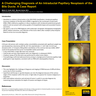
P3622 - A Challenging Endoscopic Diagnosis of an Intraductal Papillary Neoplasm of the Bile Ducts: A Case Report
Tuesday, October 29, 2024
10:30 AM - 4:00 PM ET
Location: Exhibit Hall E

Has Audio

Melis G. Celdir, MD
University of Iowa Hospitals & Clinics
Iowa City, IA
Presenting Author(s)
Melis G.. Celdir, MD, Munish Ashat, MD
University of Iowa Hospitals & Clinics, Iowa City, IA
Introduction: Intraductal papillary neoplasm of the bile duct (IPNB) is a rare disease encompassing intraductal papillary cholangiocarcinoma and precursor biliary intraductal papillary neoplasm. It is not well characterized; clinical features are described based on small case series. We report a case of an IPNB diagnosis with endoscopic retrograde cholangiopancreatography (ERCP) with balloon extraction and collection of the tumor debris after multiple sessions of cholangiography and cholangioscopy with biopsies failed to obtain the tumor tissue.
Case Description/Methods: A 94-year-old woman with multiple cardiac comorbidities presented with worsening fatigue and 45 lbs. of unintentional weight loss over 18 months. Laboratory studies revealed elevated liver chemistries: AST 82, ALT 153, total bilirubin 1.7, ALP 146. Abdominal ultrasound and CT showed a filling defect in the common bile duct (CBD) with dilation concerning a mass or stone. MRCP could not be performed due to her incompatible cardiac pacemaker. A large, elongated CBD stone and sludge in bile ducts were removed in the initial ERCP. Because of persistently elevated liver chemistries and high suspicion of malignancy, the patient underwent repeat ERCPs with cholangiography and cholangioscopy. Multiple cold forceps and spy bite biopsies of a mucin-covered lesion with papillary projections invading the CBD were nondiagnostic, showing inflamed polypoid granulation tissue and inflammation in pathologic exams. In the subsequent ERCP with balloon sweep of the bile ducts, a large tumor debris with mucous was collected. Final pathology confirmed adenocarcinoma arising in a background of extensive papillary high-grade gastric-type dysplasia, consistent with an IPNB with tubulopapillary features. CA 19-9 was elevated to 821. Given the slow growth of the tumor, the patient’s age and comorbidities, the decision was made for conservative management.
Discussion: IPNB diagnosis with accurate staging is challenging because of the various stages of the neoplasm within the tumor, diffuse or multifocal disease, and difficulties in biliary access due to obstructing mucin and tumor. Superficial cold forceps or spy bite biopsies may miss the diagnosis due to granulation tissue and thick mucin covering the tumor; therefore, a high index of suspicion for possible invasive malignancy is important. ERCP with balloon extraction of tumor debris can be utilized for accurate pathologic diagnosis and evaluation of depth of invasion.

Disclosures:
Melis G.. Celdir, MD, Munish Ashat, MD. P3622 - A Challenging Endoscopic Diagnosis of an Intraductal Papillary Neoplasm of the Bile Ducts: A Case Report, ACG 2024 Annual Scientific Meeting Abstracts. Philadelphia, PA: American College of Gastroenterology.
University of Iowa Hospitals & Clinics, Iowa City, IA
Introduction: Intraductal papillary neoplasm of the bile duct (IPNB) is a rare disease encompassing intraductal papillary cholangiocarcinoma and precursor biliary intraductal papillary neoplasm. It is not well characterized; clinical features are described based on small case series. We report a case of an IPNB diagnosis with endoscopic retrograde cholangiopancreatography (ERCP) with balloon extraction and collection of the tumor debris after multiple sessions of cholangiography and cholangioscopy with biopsies failed to obtain the tumor tissue.
Case Description/Methods: A 94-year-old woman with multiple cardiac comorbidities presented with worsening fatigue and 45 lbs. of unintentional weight loss over 18 months. Laboratory studies revealed elevated liver chemistries: AST 82, ALT 153, total bilirubin 1.7, ALP 146. Abdominal ultrasound and CT showed a filling defect in the common bile duct (CBD) with dilation concerning a mass or stone. MRCP could not be performed due to her incompatible cardiac pacemaker. A large, elongated CBD stone and sludge in bile ducts were removed in the initial ERCP. Because of persistently elevated liver chemistries and high suspicion of malignancy, the patient underwent repeat ERCPs with cholangiography and cholangioscopy. Multiple cold forceps and spy bite biopsies of a mucin-covered lesion with papillary projections invading the CBD were nondiagnostic, showing inflamed polypoid granulation tissue and inflammation in pathologic exams. In the subsequent ERCP with balloon sweep of the bile ducts, a large tumor debris with mucous was collected. Final pathology confirmed adenocarcinoma arising in a background of extensive papillary high-grade gastric-type dysplasia, consistent with an IPNB with tubulopapillary features. CA 19-9 was elevated to 821. Given the slow growth of the tumor, the patient’s age and comorbidities, the decision was made for conservative management.
Discussion: IPNB diagnosis with accurate staging is challenging because of the various stages of the neoplasm within the tumor, diffuse or multifocal disease, and difficulties in biliary access due to obstructing mucin and tumor. Superficial cold forceps or spy bite biopsies may miss the diagnosis due to granulation tissue and thick mucin covering the tumor; therefore, a high index of suspicion for possible invasive malignancy is important. ERCP with balloon extraction of tumor debris can be utilized for accurate pathologic diagnosis and evaluation of depth of invasion.

Figure: A. Endoscopic retrograde cholangiography shows a filling defect in the common hepatic duct and the bile ducts are dilated.
B. Spy cholangioscopy image of the mucin-covered tumor in the common bile duct
C. Endoscopic image of the biliary tumor debris after balloon sweep of bile ducts
B. Spy cholangioscopy image of the mucin-covered tumor in the common bile duct
C. Endoscopic image of the biliary tumor debris after balloon sweep of bile ducts
Disclosures:
Melis Celdir indicated no relevant financial relationships.
Munish Ashat indicated no relevant financial relationships.
Melis G.. Celdir, MD, Munish Ashat, MD. P3622 - A Challenging Endoscopic Diagnosis of an Intraductal Papillary Neoplasm of the Bile Ducts: A Case Report, ACG 2024 Annual Scientific Meeting Abstracts. Philadelphia, PA: American College of Gastroenterology.
