Tuesday Poster Session
Category: Colon
P3704 - Disseminated Coccidioidomycosis Masquerading as Metastasis from Colon Cancer
Tuesday, October 29, 2024
10:30 AM - 4:00 PM ET
Location: Exhibit Hall E

Has Audio
- XL
Xue Jing Li, MD
University of Arizona College of Medicine, Phoenix VA Medical Center
Phoenix, AZ
Presenting Author(s)
Xue Jing Li, MD1, Andrew Bell, MS2, Darshil Shah, MD, MPH3, Shiva Ratuapli, MD4
1University of Arizona College of Medicine, Phoenix VA Medical Center, Phoenix, AZ; 2Digestive Health Institute & Advanced Liver Disease and Liver Transplant Center, Banner University Medical Center, Phoenix, AZ; 3Ironwood Cancer and Research Centers, Goodyear, AZ; 4University of Arizona College of Medicine, Phoenix VA Medical Center, Scottsdale, AZ
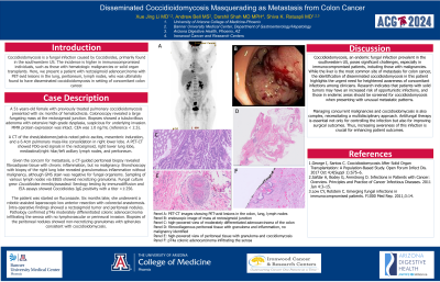
Introduction: Coccidioidomycosis is a fungal infection caused by Coccidioides, primarily found in the southwestern US. The incidence is higher in immunocompromised individuals, such as those with hematologic malignancies or solid organ transplants. We present a patient with rectosigmoid adenocarcinoma with PET-avid lesions in the lung, peritoneum, lymph nodes, who was ultimately found to have disseminated coccidioidomycosis.
Case Description/Methods: A 51-years-old female with previously treated pulmonary coccidioidomycosis presented with six months of hematochezia. Colonoscopy revealed a large fungating mass at the rectosigmoid junction. Biopsies showed a tubulovillous adenoma with extensive high-grade dysplasia, suspicious for underlying invasion. MMR protein expression was intact. CEA was 1.0 ng/mL (reference < 2.5).
A CT of the chest/abdomen/pelvis noted pelvic ascites, mesenteric induration, and a 6.4cm pulmonary mass-like consolidation in right lower lobe. A PET-CT showed FDG-avid signals in the rectosigmoid, right lower lung lobe, mediastinal/right hilar/left axillary lymph nodes, and peritoneum.
Given concern for metastasis, a CT-guided peritoneal biopsy revealed fibroadipose tissue with chronic inflammation, but no malignancy. Bronchoscopy with biopsy of the right lung lobe revealed granulomatous inflammation without malignancy, although GMS stain was negative for fungal organisms. Sampling of various lymph nodes via EBUS showed necrotizing granuloma. Fungal culture grew Coccidioides immitis/posadasii. Serology testing by immunodiffusion and EIA assays showed Coccidioides IgG positivity with a titer >1:256.
The patient was started on fluconazole. Six months later, she underwent a robotic-assisted laparoscopic low anterior resection with colorectal anastomosis. Intra-operative findings showed a rectosigmoid tumor and peritoneal nodules. Pathology confirmed pT4a moderately differentiated colonic adenocarcinoma infiltrating the serosa with no lymphovascular or perineural invasion. Biopsies of the peritoneal nodules showed non-necrotizing granulomas with spherules consistent with coccidioidomycosis.
Discussion: The most common site of metastasis for colon cancer is the liver, followed by lungs, peritoneum, and lymph nodes. This case involves a patient with newly diagnosed colon cancer with concern of metastasis who was found to have disseminated coccidioidomycosis. In endemic areas, clinicians should be vigilant about screening for coccidioidomycosis in cancer patients with abnormal metastatic patterns.

Disclosures:
Xue Jing Li, MD1, Andrew Bell, MS2, Darshil Shah, MD, MPH3, Shiva Ratuapli, MD4. P3704 - Disseminated Coccidioidomycosis Masquerading as Metastasis from Colon Cancer, ACG 2024 Annual Scientific Meeting Abstracts. Philadelphia, PA: American College of Gastroenterology.
1University of Arizona College of Medicine, Phoenix VA Medical Center, Phoenix, AZ; 2Digestive Health Institute & Advanced Liver Disease and Liver Transplant Center, Banner University Medical Center, Phoenix, AZ; 3Ironwood Cancer and Research Centers, Goodyear, AZ; 4University of Arizona College of Medicine, Phoenix VA Medical Center, Scottsdale, AZ
Introduction: Coccidioidomycosis is a fungal infection caused by Coccidioides, primarily found in the southwestern US. The incidence is higher in immunocompromised individuals, such as those with hematologic malignancies or solid organ transplants. We present a patient with rectosigmoid adenocarcinoma with PET-avid lesions in the lung, peritoneum, lymph nodes, who was ultimately found to have disseminated coccidioidomycosis.
Case Description/Methods: A 51-years-old female with previously treated pulmonary coccidioidomycosis presented with six months of hematochezia. Colonoscopy revealed a large fungating mass at the rectosigmoid junction. Biopsies showed a tubulovillous adenoma with extensive high-grade dysplasia, suspicious for underlying invasion. MMR protein expression was intact. CEA was 1.0 ng/mL (reference < 2.5).
A CT of the chest/abdomen/pelvis noted pelvic ascites, mesenteric induration, and a 6.4cm pulmonary mass-like consolidation in right lower lobe. A PET-CT showed FDG-avid signals in the rectosigmoid, right lower lung lobe, mediastinal/right hilar/left axillary lymph nodes, and peritoneum.
Given concern for metastasis, a CT-guided peritoneal biopsy revealed fibroadipose tissue with chronic inflammation, but no malignancy. Bronchoscopy with biopsy of the right lung lobe revealed granulomatous inflammation without malignancy, although GMS stain was negative for fungal organisms. Sampling of various lymph nodes via EBUS showed necrotizing granuloma. Fungal culture grew Coccidioides immitis/posadasii. Serology testing by immunodiffusion and EIA assays showed Coccidioides IgG positivity with a titer >1:256.
The patient was started on fluconazole. Six months later, she underwent a robotic-assisted laparoscopic low anterior resection with colorectal anastomosis. Intra-operative findings showed a rectosigmoid tumor and peritoneal nodules. Pathology confirmed pT4a moderately differentiated colonic adenocarcinoma infiltrating the serosa with no lymphovascular or perineural invasion. Biopsies of the peritoneal nodules showed non-necrotizing granulomas with spherules consistent with coccidioidomycosis.
Discussion: The most common site of metastasis for colon cancer is the liver, followed by lungs, peritoneum, and lymph nodes. This case involves a patient with newly diagnosed colon cancer with concern of metastasis who was found to have disseminated coccidioidomycosis. In endemic areas, clinicians should be vigilant about screening for coccidioidomycosis in cancer patients with abnormal metastatic patterns.

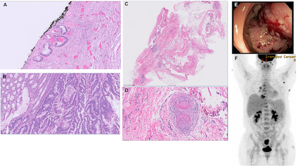
Figure: Panel A: pT4a colonic adenocarcinoma infiltrating the serosa /
Panel B: high-powered view of moderately differentiated adenocarcinoma of the colon/
Panel C: fibrocollagenous peritoneal tissue with granuloma and inflammation, no malignancy identified/
Panel D: high-powered view of peritoneal tissue with granuloma and coccidiomycosis/
Panel E: endoscopic image of mass at rectosigmoid junction/
Panel F: PET-CT images showing PET-avid lesions in the colon, lung, lymph nodes
Disclosures:
Xue Jing Li indicated no relevant financial relationships.
Andrew Bell indicated no relevant financial relationships.
Darshil Shah: Ipsen Biopharma – Advisor or Review Panel Member. Tempus Labs – Advisor or Review Panel Member.
Shiva Ratuapli indicated no relevant financial relationships.
Xue Jing Li, MD1, Andrew Bell, MS2, Darshil Shah, MD, MPH3, Shiva Ratuapli, MD4. P3704 - Disseminated Coccidioidomycosis Masquerading as Metastasis from Colon Cancer, ACG 2024 Annual Scientific Meeting Abstracts. Philadelphia, PA: American College of Gastroenterology.
