Tuesday Poster Session
Category: Colon
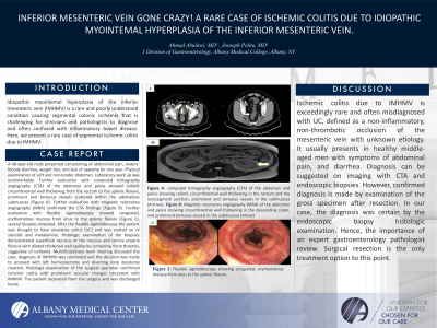
P3726 - Inferior Mesenteric Vein Gone Crazy! A Rare Case of Ischemic Colitis Due to Idiopathic Myointemal Hyperplasia of the Inferior Mesenteric Vein.
Tuesday, October 29, 2024
10:30 AM - 4:00 PM ET
Location: Exhibit Hall E


Ahmad Abulawi, MD
Albany Medical College
Albany, NY
Presenting Author(s)
Ahmad Abulawi, MD, Joseph Polito, MD
Albany Medical College, Albany, NY
Introduction: Idiopathic myointemal hyperplasia of the inferior mesenteric vein (IMHMV) is a rare and poorly understood condition causing segmental colonic ischemia that is challenging for clinicians and pathologists to diagnose and often confused with inflammatory bowel disease. Here, we present a rare case of segmental ischemic colitis due to IMHMV.
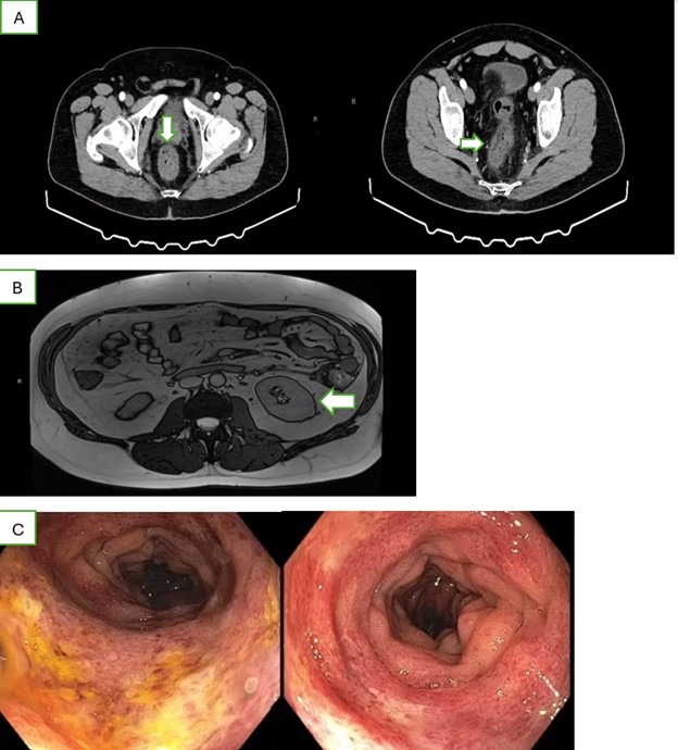
Case Description/Methods: A 48-year-old male with no past medical history presented complaining of abdominal pain, watery-bloody diarrhea, weight loss, and lack of appetite for one year. Physical examination of soft and non-tender abdomen. Laboratory work up was unremarkable. Further evaluation with computed tomography angiography (CTA) of the abdomen and pelvis showed colonic circumferential wall thickening from the rectum to the splenic flexure, prominent and tortuous vessels scattered within the edematous submucosa (Figure A). Further evaluation with magnetic resonance angiography (MRA) confirmed the CTA findings (Figure B). Further evaluation with flexible sigmoidoscopy showed congested, erythematous mucosa from anus to the splenic flexure (Figure C), several biopsies obtained. After the flexible sigmoidoscopy the patient was thought to have ulcerative colitis (UC) and was started on IV steroids and mesalamine. Histologic examination of the biopsies demonstrated superficial necrosis of the mucosa and lamina propria fibrosis with dilated thickened wall capillaries containing fibrin thrombi, suggestive of ischemia. Multidisciplinary team meeting discussed the case, diagnosis of IMHMV was concluded and the decision was made to proceed with left hemicolectomy and diverting loop ileostomy creation. Histologic examination of the surgical specimen confirmed ischemic colitis with prominent vascular changes consistent with IMHMV. The patient recovered from the surgery and was discharged home.
Discussion: Ischemic colitis due to IMHMV is exceedingly rare and often misdiagnosed with UC, defined as a non-inflammatory, non-thrombotic occlusion of the mesenteric vein with unknown etiology. It usually presents in healthy middle-aged men with symptoms of abdominal pain, and diarrhea. Diagnosis can be suggested on imaging with CTA and endoscopic biopsies. However, confirmed diagnosis is made by examination of the gross specimen after resection. In our case, the diagnosis was certain by the endoscopic biopsy histologic examination. Hence, the importance of an expert gastroenterology pathologist review. Surgical resection is the only treatment option to this point.

Disclosures:
Ahmad Abulawi, MD, Joseph Polito, MD. P3726 - Inferior Mesenteric Vein Gone Crazy! A Rare Case of Ischemic Colitis Due to Idiopathic Myointemal Hyperplasia of the Inferior Mesenteric Vein., ACG 2024 Annual Scientific Meeting Abstracts. Philadelphia, PA: American College of Gastroenterology.
Albany Medical College, Albany, NY
Introduction: Idiopathic myointemal hyperplasia of the inferior mesenteric vein (IMHMV) is a rare and poorly understood condition causing segmental colonic ischemia that is challenging for clinicians and pathologists to diagnose and often confused with inflammatory bowel disease. Here, we present a rare case of segmental ischemic colitis due to IMHMV.
Case Description/Methods: A 48-year-old male with no past medical history presented complaining of abdominal pain, watery-bloody diarrhea, weight loss, and lack of appetite for one year. Physical examination of soft and non-tender abdomen. Laboratory work up was unremarkable. Further evaluation with computed tomography angiography (CTA) of the abdomen and pelvis showed colonic circumferential wall thickening from the rectum to the splenic flexure, prominent and tortuous vessels scattered within the edematous submucosa (Figure A). Further evaluation with magnetic resonance angiography (MRA) confirmed the CTA findings (Figure B). Further evaluation with flexible sigmoidoscopy showed congested, erythematous mucosa from anus to the splenic flexure (Figure C), several biopsies obtained. After the flexible sigmoidoscopy the patient was thought to have ulcerative colitis (UC) and was started on IV steroids and mesalamine. Histologic examination of the biopsies demonstrated superficial necrosis of the mucosa and lamina propria fibrosis with dilated thickened wall capillaries containing fibrin thrombi, suggestive of ischemia. Multidisciplinary team meeting discussed the case, diagnosis of IMHMV was concluded and the decision was made to proceed with left hemicolectomy and diverting loop ileostomy creation. Histologic examination of the surgical specimen confirmed ischemic colitis with prominent vascular changes consistent with IMHMV. The patient recovered from the surgery and was discharged home.
Discussion: Ischemic colitis due to IMHMV is exceedingly rare and often misdiagnosed with UC, defined as a non-inflammatory, non-thrombotic occlusion of the mesenteric vein with unknown etiology. It usually presents in healthy middle-aged men with symptoms of abdominal pain, and diarrhea. Diagnosis can be suggested on imaging with CTA and endoscopic biopsies. However, confirmed diagnosis is made by examination of the gross specimen after resection. In our case, the diagnosis was certain by the endoscopic biopsy histologic examination. Hence, the importance of an expert gastroenterology pathologist review. Surgical resection is the only treatment option to this point.

Figure: Figure A: computed tomography angiography (CTA) of the abdomen and pelvis showing colonic circumferential wall thickening in the rectum and the rectosigmoid junction, prominent and tortuous vessels in the submucosa (Arrows).
Figure B: Magnetic resonance angiography (MRA) of the abdomen and pelvis showing circumferential wall thickening in the descending colon, and prominent tortuous vessels in the submucosa (Arrow).
Figure C: Flexible sigmoidoscopy showing congested, erythematous mucosa from anus to the splenic flexure.
Disclosures:
Ahmad Abulawi indicated no relevant financial relationships.
Joseph Polito indicated no relevant financial relationships.
Ahmad Abulawi, MD, Joseph Polito, MD. P3726 - Inferior Mesenteric Vein Gone Crazy! A Rare Case of Ischemic Colitis Due to Idiopathic Myointemal Hyperplasia of the Inferior Mesenteric Vein., ACG 2024 Annual Scientific Meeting Abstracts. Philadelphia, PA: American College of Gastroenterology.
