Tuesday Poster Session
Category: Colon
P3738 - What Exactly Is Pushing the Cecum? A Diagnostic Challenge
Tuesday, October 29, 2024
10:30 AM - 4:00 PM ET
Location: Exhibit Hall E

Has Audio

Luis Biescas-Gonzalez, MD
VA Caribbean Healthcare System
San Juan, PR
Presenting Author(s)
Luis Biescas-Gonzalez, MD, Ricardo Lopez-Valle, MD, Jose Sorrentino-Brunisholz, MD, Juan Diaz-Hernandez, MD, Andres Rabell-Bernal, MD
VA Caribbean Healthcare System, San Juan, Puerto Rico
Introduction: Although colonoscopies are probably best known as tools for colorectal cancer screening, we should never diminish their importance in diagnosing other conditions associated with the gastrointestinal (GI) tract. During a screening colonoscopy we may often stumble upon some rather uncommon findings, and some of these findings may not even be inside the GI tract. In the case below we highlight a rare presentation of an extracolonic lesion identified during a colonoscopy and how extensive exploration was needed to arrive at a correct diagnosis.
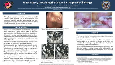
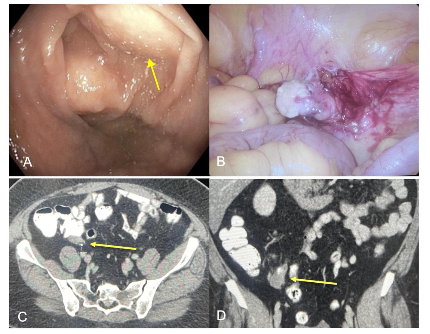
Case Description/Methods: A 56-year-old female presented to the GI unit for a routine screening colonoscopy, she was asymptomatic, with regular bowel movements, and no concerning alarm signs or symptoms. Colonoscopy revealed what appeared to be a subepithelial lesion adjacent to the appendiceal orifice without other concerning mucosal abnormalities (Fig 1). Differential diagnoses included an external structure causing a mass effect upon the cecum or an appendiceal mucocele. An Abdominopelvic CT was requested revealing a smooth, partially calcified, hypodense 3 cm soft tissue lesion abutting vs. exophytically arising from the posterior surface of the cecum, separate from the appendix (Fig 1). Due to its location, a percutaneous biopsy was deemed risky by the interventional radiology service since the lesion was mobile and proximity to the bowel could lead to perforation. A diagnostic laparoscopy was performed and revealed a mass near the cecum, adhered to the abdominal wall. The OB-Gyn service was consulted intra-op and confirmed the mass was a calcified ovary which was consistent with the patient's history of a unilateral salpingo-oophorectomy. The ovary was left in place, and she was discharged home with an uneventful postoperative recovery.
Discussion: This case denotes the diagnostic challenges that may arise from an incidental endoscopic finding. An adhered ovary protruding into the cecum, was misinterpreted as an exophytic cecal mass, underscoring the necessity of considering unexpected differential diagnoses in patients with unclear surgical histories. A few similar presentations have been described in the literature, including a dominant ovarian follicle and a right hernia repair plug causing extrinsic compression of the cecal wall1. Recognition of these rare presentations can aid clinicians in considering similar differential diagnoses in future cases.
References:
1. Mudaliar S, et al. Ovary unusual colonic lesion. JGH Open. 2021 Jun 2;5(7):834-836.

Disclosures:
Luis Biescas-Gonzalez, MD, Ricardo Lopez-Valle, MD, Jose Sorrentino-Brunisholz, MD, Juan Diaz-Hernandez, MD, Andres Rabell-Bernal, MD. P3738 - What Exactly Is Pushing the Cecum? A Diagnostic Challenge, ACG 2024 Annual Scientific Meeting Abstracts. Philadelphia, PA: American College of Gastroenterology.
VA Caribbean Healthcare System, San Juan, Puerto Rico
Introduction: Although colonoscopies are probably best known as tools for colorectal cancer screening, we should never diminish their importance in diagnosing other conditions associated with the gastrointestinal (GI) tract. During a screening colonoscopy we may often stumble upon some rather uncommon findings, and some of these findings may not even be inside the GI tract. In the case below we highlight a rare presentation of an extracolonic lesion identified during a colonoscopy and how extensive exploration was needed to arrive at a correct diagnosis.
Case Description/Methods: A 56-year-old female presented to the GI unit for a routine screening colonoscopy, she was asymptomatic, with regular bowel movements, and no concerning alarm signs or symptoms. Colonoscopy revealed what appeared to be a subepithelial lesion adjacent to the appendiceal orifice without other concerning mucosal abnormalities (Fig 1). Differential diagnoses included an external structure causing a mass effect upon the cecum or an appendiceal mucocele. An Abdominopelvic CT was requested revealing a smooth, partially calcified, hypodense 3 cm soft tissue lesion abutting vs. exophytically arising from the posterior surface of the cecum, separate from the appendix (Fig 1). Due to its location, a percutaneous biopsy was deemed risky by the interventional radiology service since the lesion was mobile and proximity to the bowel could lead to perforation. A diagnostic laparoscopy was performed and revealed a mass near the cecum, adhered to the abdominal wall. The OB-Gyn service was consulted intra-op and confirmed the mass was a calcified ovary which was consistent with the patient's history of a unilateral salpingo-oophorectomy. The ovary was left in place, and she was discharged home with an uneventful postoperative recovery.
Discussion: This case denotes the diagnostic challenges that may arise from an incidental endoscopic finding. An adhered ovary protruding into the cecum, was misinterpreted as an exophytic cecal mass, underscoring the necessity of considering unexpected differential diagnoses in patients with unclear surgical histories. A few similar presentations have been described in the literature, including a dominant ovarian follicle and a right hernia repair plug causing extrinsic compression of the cecal wall1. Recognition of these rare presentations can aid clinicians in considering similar differential diagnoses in future cases.
References:
1. Mudaliar S, et al. Ovary unusual colonic lesion. JGH Open. 2021 Jun 2;5(7):834-836.

Figure: Figure 1: (A-B) Colonoscopy view of the cecum; a broad-based mass is present. The mucosa appears normal and did not exhibit erythema or ulcerations. Abdominopelvic CT with IV contrast (C) axial and (D) coronal views demonstrating an oval-shaped hypodense mass abutting the cecum or exophytically arising from the posterior serosal surface of the cecum at the level of the appendiceal orifice. The appendix is normal in caliber and appears to be separate from the lesion.
Disclosures:
Luis Biescas-Gonzalez indicated no relevant financial relationships.
Ricardo Lopez-Valle indicated no relevant financial relationships.
Jose Sorrentino-Brunisholz indicated no relevant financial relationships.
Juan Diaz-Hernandez indicated no relevant financial relationships.
Andres Rabell-Bernal indicated no relevant financial relationships.
Luis Biescas-Gonzalez, MD, Ricardo Lopez-Valle, MD, Jose Sorrentino-Brunisholz, MD, Juan Diaz-Hernandez, MD, Andres Rabell-Bernal, MD. P3738 - What Exactly Is Pushing the Cecum? A Diagnostic Challenge, ACG 2024 Annual Scientific Meeting Abstracts. Philadelphia, PA: American College of Gastroenterology.
