Tuesday Poster Session
Category: Colon
P3739 - A Case of Colonic Inflammatory Myofibroblastic Tumor
Tuesday, October 29, 2024
10:30 AM - 4:00 PM ET
Location: Exhibit Hall E

Has Audio

Mohammad Sultan, DO
Ochsner Health Clinic Foundation
New Orleans, LA
Presenting Author(s)
Mohammad Sultan, DO, Neej Patel, MD
Ochsner Health Clinic Foundation, New Orleans, LA
Introduction: Sarcomas are tumors of connective tissue that arise from mesenchymal tissue. Uncommon subtypes (including Inflammatory Myofibroblastic Tumors or IMT's) can be challenging to diagnose and treat given they account for less than 5 percent of all sarcomas.1 IMT's, which are benign, tend to be found in children with a mean age of 9-10.2 Histology is the gold standard for diagnosis and while there are no known risk factors for developing IMT’s, there are certain characteristics worth noting. We discuss a case of a patient referred to our facility for Endoscopic Ultrasound (EUS) of a sigmoid mass.
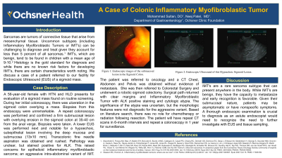
Case Description/Methods: A 56-year-old female with HTN and HLD presents for evaluation of a sigmoid mass. During her initial colonoscopy, there was ulceration in the sigmoid colon overlying a mass. Biopsies from this endoscopy were unremarkable. A repeat colonoscopy was performed and confirmed a firm submucosal lesion with overlying erosion in the sigmoid colon at 35-40 cm from the anal verge. Biopsies were taken. A lower EUS was performed and notable for a hypoechoic, subepithelial lesion involving the deep mucosa and submucosa (13x10 mm). Transcolonic fine needle aspiration was obtained. Pathology was unclear, but the tumor stained positive for ALK. This raised concerns for epithelioid inflammatory myofibroblastic sarcoma; an aggressive intra-abdominal variant of IMT.
The patient was referred to oncology and a CT Chest, Abdomen and Pelvis was obtained without evidence of metastasis. She was then referred to Colorectal Surgery and underwent a robotic sigmoid colectomy. Surgical path returned with clear margins and Inflammatory Myofibroblastic Tumor with ALK positive staining and cytologic atypia. The significance of the atypia was uncertain, but the morphologic features were not diagnostic for the aggressive variant. Based on literature review, there was no role for chemotherapy or radiation following resection. The patient will have repeat CT scans in 6-month intervals and repeat a colonoscopy in 1 year for surveillance.
Discussion: IMT's are a rare sarcoma subtype that can present anywhere in the body. While IMT’s are benign, they have the capacity to metastasize and early recognition is favorable. Given their submucosal nature, patients may be asymptomatic or have nonspecific symptoms. A thorough endoscopic examination is crucial to diagnosis as an astute endoscopist would need to recognize the need to further investigate with EUS and tissue sampling.

Disclosures:
Mohammad Sultan, DO, Neej Patel, MD. P3739 - A Case of Colonic Inflammatory Myofibroblastic Tumor, ACG 2024 Annual Scientific Meeting Abstracts. Philadelphia, PA: American College of Gastroenterology.
Ochsner Health Clinic Foundation, New Orleans, LA
Introduction: Sarcomas are tumors of connective tissue that arise from mesenchymal tissue. Uncommon subtypes (including Inflammatory Myofibroblastic Tumors or IMT's) can be challenging to diagnose and treat given they account for less than 5 percent of all sarcomas.1 IMT's, which are benign, tend to be found in children with a mean age of 9-10.2 Histology is the gold standard for diagnosis and while there are no known risk factors for developing IMT’s, there are certain characteristics worth noting. We discuss a case of a patient referred to our facility for Endoscopic Ultrasound (EUS) of a sigmoid mass.
Case Description/Methods: A 56-year-old female with HTN and HLD presents for evaluation of a sigmoid mass. During her initial colonoscopy, there was ulceration in the sigmoid colon overlying a mass. Biopsies from this endoscopy were unremarkable. A repeat colonoscopy was performed and confirmed a firm submucosal lesion with overlying erosion in the sigmoid colon at 35-40 cm from the anal verge. Biopsies were taken. A lower EUS was performed and notable for a hypoechoic, subepithelial lesion involving the deep mucosa and submucosa (13x10 mm). Transcolonic fine needle aspiration was obtained. Pathology was unclear, but the tumor stained positive for ALK. This raised concerns for epithelioid inflammatory myofibroblastic sarcoma; an aggressive intra-abdominal variant of IMT.
The patient was referred to oncology and a CT Chest, Abdomen and Pelvis was obtained without evidence of metastasis. She was then referred to Colorectal Surgery and underwent a robotic sigmoid colectomy. Surgical path returned with clear margins and Inflammatory Myofibroblastic Tumor with ALK positive staining and cytologic atypia. The significance of the atypia was uncertain, but the morphologic features were not diagnostic for the aggressive variant. Based on literature review, there was no role for chemotherapy or radiation following resection. The patient will have repeat CT scans in 6-month intervals and repeat a colonoscopy in 1 year for surveillance.
Discussion: IMT's are a rare sarcoma subtype that can present anywhere in the body. While IMT’s are benign, they have the capacity to metastasize and early recognition is favorable. Given their submucosal nature, patients may be asymptomatic or have nonspecific symptoms. A thorough endoscopic examination is crucial to diagnosis as an astute endoscopist would need to recognize the need to further investigate with EUS and tissue sampling.

Figure: Figure 1: Endoscopic Images of Sigmoid Subepithelial Mass
Disclosures:
Mohammad Sultan indicated no relevant financial relationships.
Neej Patel indicated no relevant financial relationships.
Mohammad Sultan, DO, Neej Patel, MD. P3739 - A Case of Colonic Inflammatory Myofibroblastic Tumor, ACG 2024 Annual Scientific Meeting Abstracts. Philadelphia, PA: American College of Gastroenterology.
