Tuesday Poster Session
Category: Colon
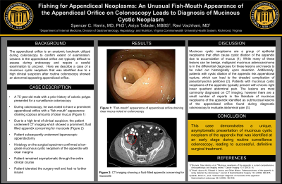
P3742 - Fishing for Appendiceal Neoplasms: An Unusual Fish-Mouth Appearance of the Appendiceal Orifice on Colonoscopy Leads to Diagnosis of Mucinous Cystic Neoplasm
Tuesday, October 29, 2024
10:30 AM - 4:00 PM ET
Location: Exhibit Hall E

Has Audio
- SH
Spencer Harris, MD, PhD
Virginia Commonwealth University Health System
Richmond, VA
Presenting Author(s)
Spencer Harris, MD, PhD, Asiya Tafader, MBBS, Ravi Vachhani, MD
Virginia Commonwealth University Health System, Richmond, VA
Introduction: Mucinous cystic neoplasms are a group of epithelial neoplasms that often cause cystic dilation of the appendix due to accumulation of mucus. While many of these lesions can be benign, malignant mucinous adenocarcinoma is in the differential diagnoses for these lesions and needs to be ruled out histologically upon resection. Additionally, patients with cystic dilation of the appendix risk appendiceal rupture, which can lead to the dreaded complication of pseudomyxoma peritonei. Typically these lesions are noted on computed tomography obtained due to abdominal pain, however here we present a case of an asymptomatic patient undergoing surveillance colonoscopy who was found to have a mucinous cystic neoplasm of the appendix.
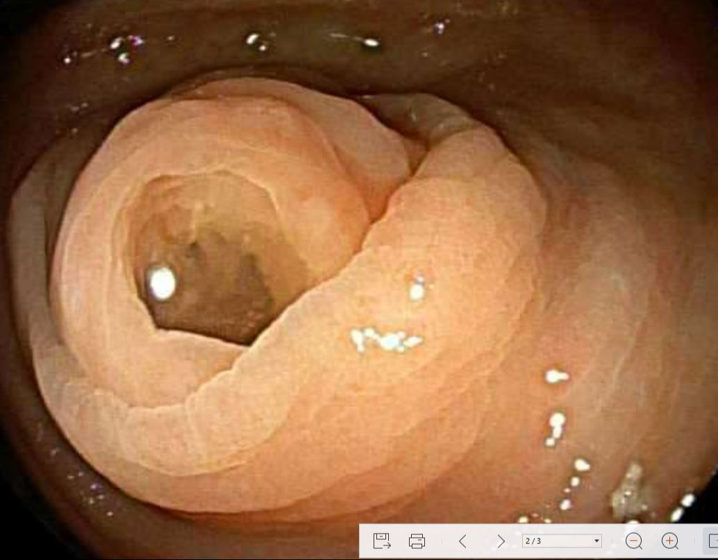
Case Description/Methods: A 75 year old male patient with medical history significant for prior colonic polyps presented for a routine surveillance colonoscopy. During his colonoscopy, he was noted to have a prominent appendiceal orifice draining a copious amount of clear mucus, with a “fish-mouth” appearance on endoscopic visual inspection. While no mucosal or submucosal abnormalities were noted on exam, the area was biopsied, but initial biopsy results showed no histopathologic abnormalities. Due to a high clinical suspicion, the patient underwent CT imaging that showed a prominent, fluid-filled appendix, concerning for a mucocele. The patient subsequently underwent laparoscopic appendectomy, and the surgical specimen confirmed a low-grade mucinous cystic neoplasm of the appendix with clear margins.
Discussion: Patients with mucinous cystic neoplasms of the appendix typically present with chronic right lower quadrant abdominal pain. The lesions are most commonly diagnosed on computed tomography imaging, however there are a small number of reports in the literature of mucinous neoplasms of the appendix identified as submucosal lesions of the appendiceal orifice found during diagnostic colonoscopy to evaluate abdominal pain. This case demonstrates a unique, asymptomatic presentation of mucinous cystic neoplasm of the appendix that was identified at an early stage during routine surveillance colonoscopy, leading to successful, definitive surgical treatment.

Disclosures:
Spencer Harris, MD, PhD, Asiya Tafader, MBBS, Ravi Vachhani, MD. P3742 - Fishing for Appendiceal Neoplasms: An Unusual Fish-Mouth Appearance of the Appendiceal Orifice on Colonoscopy Leads to Diagnosis of Mucinous Cystic Neoplasm, ACG 2024 Annual Scientific Meeting Abstracts. Philadelphia, PA: American College of Gastroenterology.
Virginia Commonwealth University Health System, Richmond, VA
Introduction: Mucinous cystic neoplasms are a group of epithelial neoplasms that often cause cystic dilation of the appendix due to accumulation of mucus. While many of these lesions can be benign, malignant mucinous adenocarcinoma is in the differential diagnoses for these lesions and needs to be ruled out histologically upon resection. Additionally, patients with cystic dilation of the appendix risk appendiceal rupture, which can lead to the dreaded complication of pseudomyxoma peritonei. Typically these lesions are noted on computed tomography obtained due to abdominal pain, however here we present a case of an asymptomatic patient undergoing surveillance colonoscopy who was found to have a mucinous cystic neoplasm of the appendix.
Case Description/Methods: A 75 year old male patient with medical history significant for prior colonic polyps presented for a routine surveillance colonoscopy. During his colonoscopy, he was noted to have a prominent appendiceal orifice draining a copious amount of clear mucus, with a “fish-mouth” appearance on endoscopic visual inspection. While no mucosal or submucosal abnormalities were noted on exam, the area was biopsied, but initial biopsy results showed no histopathologic abnormalities. Due to a high clinical suspicion, the patient underwent CT imaging that showed a prominent, fluid-filled appendix, concerning for a mucocele. The patient subsequently underwent laparoscopic appendectomy, and the surgical specimen confirmed a low-grade mucinous cystic neoplasm of the appendix with clear margins.
Discussion: Patients with mucinous cystic neoplasms of the appendix typically present with chronic right lower quadrant abdominal pain. The lesions are most commonly diagnosed on computed tomography imaging, however there are a small number of reports in the literature of mucinous neoplasms of the appendix identified as submucosal lesions of the appendiceal orifice found during diagnostic colonoscopy to evaluate abdominal pain. This case demonstrates a unique, asymptomatic presentation of mucinous cystic neoplasm of the appendix that was identified at an early stage during routine surveillance colonoscopy, leading to successful, definitive surgical treatment.

Figure: ”Fish-mouth” appearance of appendiceal orifice draining clear mucus noted on colonoscopy
Disclosures:
Spencer Harris indicated no relevant financial relationships.
Asiya Tafader indicated no relevant financial relationships.
Ravi Vachhani indicated no relevant financial relationships.
Spencer Harris, MD, PhD, Asiya Tafader, MBBS, Ravi Vachhani, MD. P3742 - Fishing for Appendiceal Neoplasms: An Unusual Fish-Mouth Appearance of the Appendiceal Orifice on Colonoscopy Leads to Diagnosis of Mucinous Cystic Neoplasm, ACG 2024 Annual Scientific Meeting Abstracts. Philadelphia, PA: American College of Gastroenterology.
