Tuesday Poster Session
Category: Colon
P3745 - A Case of Atypical Endometriosis Presenting as a Malignant Appearing Colonic Mass
Tuesday, October 29, 2024
10:30 AM - 4:00 PM ET
Location: Exhibit Hall E

Has Audio
- AT
Akanksha Togra, MD
Texas Tech University Health Sciences Center
El Paso, Texas
Presenting Author(s)
Award: Presidential Poster Award
Akanksha Togra, MD, Swati Mahapatra, DO, M Ammar Kalas, MD, Keith Garrison, MD, Alejandro Robles, MD
Texas Tech University Health Sciences Center, El Paso, TX
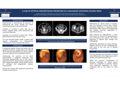
Introduction: Endometriosis is a chronic gynecological condition characterized by presence of endometrial-like tissue outside the uterus, leading to pelvic pain and infertility. It typically presents in pelvic sites such as the ovaries (most common), fallopian tubes, pelvic peritoneum and uterosacral ligaments. Atypically, it can present in extra pelvic sites such as the urinary tract, thorax, gastrointestinal tract, and soft tissues. Herein, we present an atypical presentation of endometriosis manifesting as chronic gastrointestinal bleed with symptomatic anemia.
Case Description/Methods: 50-year-old female was referred from her primary care physician in Mexico due to symptoms of fatigue secondary to severe anemia with hemoglobin of 4mg/dL. Gastroenterology was consulted for evaluation of anemia. EGD was performed and was unremarkable. Colonoscopy revealed a malignant appearing mass measuring approximately 3cm in the cecum/appendiceal orifice. A biopsy of the cecal/appendiceal orifice lesion showed granulation tissue and endometriosis with entrapped hyperplastic colonic glands. Endometrial glands and stroma were positive for estrogen receptors and progesterone receptor immunostains. Due to the endoscopic findings and significant anemia, surgery was consulted and patient underwent exploratory laparotomy for right hemicolectomy.
Discussion: Colonic lesions noted in patients with iron deficiency anemia often indicate underlying malignancy or advanced polyps. Appendiceal orifice lesions could indicate underlying mucocele, appendicitis/appendiceal abscess, or mucinous appendiceal neoplasms.
The symptomatology and physical findings in cecal diseases tend to be less diagnostic than other parts of the gastrointestinal tract. Additionally, a variety of lesions may simulate colonic neoplasms and it’s imperative to differentiate them from true gastrointestinal neoplasms. With reference to endometriosis, a mere 0.3% of these cases are located in the gastrointestinal tract and even lesser in the cecum. These factors make diagnosis of endometriosis at extraovarian locations such as the cecum challenging, both clinically and radiologically.
This case illustrates the importance of having a broad differentials when diagnosing anemic patients with gastrointestinal mass. While not common, consideration of alternate etiologies for colonic lesions should be considered in patients without clear endoscopic appearance of colonic malignancy and no risk factors for colon cancer.

Disclosures:
Akanksha Togra, MD, Swati Mahapatra, DO, M Ammar Kalas, MD, Keith Garrison, MD, Alejandro Robles, MD. P3745 - A Case of Atypical Endometriosis Presenting as a Malignant Appearing Colonic Mass, ACG 2024 Annual Scientific Meeting Abstracts. Philadelphia, PA: American College of Gastroenterology.
Akanksha Togra, MD, Swati Mahapatra, DO, M Ammar Kalas, MD, Keith Garrison, MD, Alejandro Robles, MD
Texas Tech University Health Sciences Center, El Paso, TX
Introduction: Endometriosis is a chronic gynecological condition characterized by presence of endometrial-like tissue outside the uterus, leading to pelvic pain and infertility. It typically presents in pelvic sites such as the ovaries (most common), fallopian tubes, pelvic peritoneum and uterosacral ligaments. Atypically, it can present in extra pelvic sites such as the urinary tract, thorax, gastrointestinal tract, and soft tissues. Herein, we present an atypical presentation of endometriosis manifesting as chronic gastrointestinal bleed with symptomatic anemia.
Case Description/Methods: 50-year-old female was referred from her primary care physician in Mexico due to symptoms of fatigue secondary to severe anemia with hemoglobin of 4mg/dL. Gastroenterology was consulted for evaluation of anemia. EGD was performed and was unremarkable. Colonoscopy revealed a malignant appearing mass measuring approximately 3cm in the cecum/appendiceal orifice. A biopsy of the cecal/appendiceal orifice lesion showed granulation tissue and endometriosis with entrapped hyperplastic colonic glands. Endometrial glands and stroma were positive for estrogen receptors and progesterone receptor immunostains. Due to the endoscopic findings and significant anemia, surgery was consulted and patient underwent exploratory laparotomy for right hemicolectomy.
Discussion: Colonic lesions noted in patients with iron deficiency anemia often indicate underlying malignancy or advanced polyps. Appendiceal orifice lesions could indicate underlying mucocele, appendicitis/appendiceal abscess, or mucinous appendiceal neoplasms.
The symptomatology and physical findings in cecal diseases tend to be less diagnostic than other parts of the gastrointestinal tract. Additionally, a variety of lesions may simulate colonic neoplasms and it’s imperative to differentiate them from true gastrointestinal neoplasms. With reference to endometriosis, a mere 0.3% of these cases are located in the gastrointestinal tract and even lesser in the cecum. These factors make diagnosis of endometriosis at extraovarian locations such as the cecum challenging, both clinically and radiologically.
This case illustrates the importance of having a broad differentials when diagnosing anemic patients with gastrointestinal mass. While not common, consideration of alternate etiologies for colonic lesions should be considered in patients without clear endoscopic appearance of colonic malignancy and no risk factors for colon cancer.

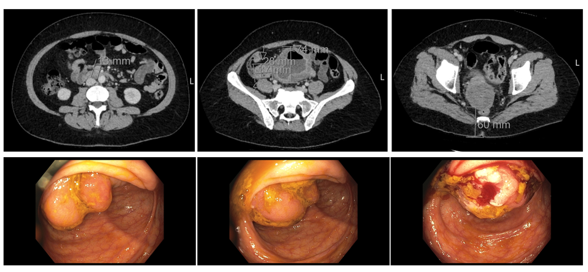
Figure: Image 1 (a, b, c): CT was concerning for appendiceal neoplastic solid lesion extending into the cecum and regional lymphadenopathy.
Image 2 (a, b, c): Colonoscopy was concerning for a 3cm malignant appearing tumor at the appendiceal orifice
Image 2 (a, b, c): Colonoscopy was concerning for a 3cm malignant appearing tumor at the appendiceal orifice
Disclosures:
Akanksha Togra: Clinexel Inc – Owner/Ownership Interest. Clinexel Life Sciences Pvt Ltd – Owner/Ownership Interest. Cytenet Life science LLP – Owner/Ownership Interest. GLRK Healthcare foundation (Non-profit Organization Company) – Owner/Ownership Interest.
Swati Mahapatra indicated no relevant financial relationships.
M Ammar Kalas indicated no relevant financial relationships.
Keith Garrison indicated no relevant financial relationships.
Alejandro Robles indicated no relevant financial relationships.
Akanksha Togra, MD, Swati Mahapatra, DO, M Ammar Kalas, MD, Keith Garrison, MD, Alejandro Robles, MD. P3745 - A Case of Atypical Endometriosis Presenting as a Malignant Appearing Colonic Mass, ACG 2024 Annual Scientific Meeting Abstracts. Philadelphia, PA: American College of Gastroenterology.

