Tuesday Poster Session
Category: Colon
P3749 - A Rare Case of Concurrent Anorectal Melanoma and Sigmoid Adenocarcinoma
Tuesday, October 29, 2024
10:30 AM - 4:00 PM ET
Location: Exhibit Hall E

Has Audio

Navin Naik, DO
Franciscan Health Olympia Fields
Tinley Park, IL
Presenting Author(s)
Navin Naik, DO1, Shoba Mendu, MD2
1Franciscan Health Olympia Fields, Tinley Park, IL; 2Gastroenterology Associates, Chesapeake, VA
Introduction: Anorectal melanoma is a rare and aggressive cancer. It accounts for less than 1% of all melanomas and less than 3% of all anorectal cancers. Prognosis is generally poor as the tumor has usually locally invaded by the time of diagnosis. Multiple primary colorectal cancers are also rare. Concurrent primary anorectal melanoma and primary colorectal adenocarcinoma is extremely rare, with only a handful of cases reported in the literature. We present a case of synchronous anorectal melanoma and sigmoid adenocarcinoma.
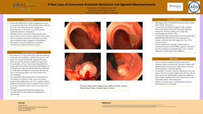
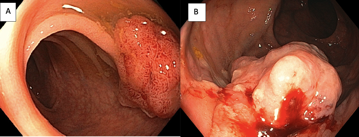
Case Description/Methods: Our patient is a 71 year old woman who was referred to the clinic for evaluation of blood and mucus in her stool. Her symptoms had been ongoing for a year before arrival, but she was unable to be seen prior. On evaluation, she was reporting seeing blood and mucus with every bowel movement and having to 2-3 movements per day. She did not have any abdominal or rectal pain, constipation or diarrhea. She had not had a colonoscopy before. Her family history was unremarkable. She underwent colonoscopy which showed two 3-4 mm polyps in transverse colon, one 4 mm sessile polyp in descending colon, one 5-6 cm pedunculated polyp with ulcerated surface in sigmoid colon (Image A), and a 2 cm friable, ulcerated mass in the rectum which was biopsied (Image B). The sigmoid polyp and remaining polyps were removed with hot and cold snare polypectomy, respectively. Pathology results showed the transverse colon polyps were tubular adenomas. The rectal mass showed malignant cells; multiple stains were performed which indicated malignant melanoma. The descending colon polyp was morphologically similar to this. The sigmoid colon polyp showed invasive, moderately differentiated adenocarcinoma arising within a tubular adenoma; the stalk margin was free of the invasive tumor. She was referred to oncology. Further work up revealed primary anorectal BRAF-negative melanoma. She was started on immunotherapy and is currently doing well 3 years after initial diagnosis.
Discussion: There are only a handful of similar cases in the literature. Most presented late and were treated with abdominoperineal resection, which seems to be the best treatment in advanced disease. Our patient is unique in that her adenocarcinoma was able to be removed with polypectomy and she did not have recurrent. Additionally, surgery was deferred due to likely metastasis from her melanoma, but she responded well to immunotherapy. Prognosis in these cases is still generally very poor and treatment is challenging.

Disclosures:
Navin Naik, DO1, Shoba Mendu, MD2. P3749 - A Rare Case of Concurrent Anorectal Melanoma and Sigmoid Adenocarcinoma, ACG 2024 Annual Scientific Meeting Abstracts. Philadelphia, PA: American College of Gastroenterology.
1Franciscan Health Olympia Fields, Tinley Park, IL; 2Gastroenterology Associates, Chesapeake, VA
Introduction: Anorectal melanoma is a rare and aggressive cancer. It accounts for less than 1% of all melanomas and less than 3% of all anorectal cancers. Prognosis is generally poor as the tumor has usually locally invaded by the time of diagnosis. Multiple primary colorectal cancers are also rare. Concurrent primary anorectal melanoma and primary colorectal adenocarcinoma is extremely rare, with only a handful of cases reported in the literature. We present a case of synchronous anorectal melanoma and sigmoid adenocarcinoma.
Case Description/Methods: Our patient is a 71 year old woman who was referred to the clinic for evaluation of blood and mucus in her stool. Her symptoms had been ongoing for a year before arrival, but she was unable to be seen prior. On evaluation, she was reporting seeing blood and mucus with every bowel movement and having to 2-3 movements per day. She did not have any abdominal or rectal pain, constipation or diarrhea. She had not had a colonoscopy before. Her family history was unremarkable. She underwent colonoscopy which showed two 3-4 mm polyps in transverse colon, one 4 mm sessile polyp in descending colon, one 5-6 cm pedunculated polyp with ulcerated surface in sigmoid colon (Image A), and a 2 cm friable, ulcerated mass in the rectum which was biopsied (Image B). The sigmoid polyp and remaining polyps were removed with hot and cold snare polypectomy, respectively. Pathology results showed the transverse colon polyps were tubular adenomas. The rectal mass showed malignant cells; multiple stains were performed which indicated malignant melanoma. The descending colon polyp was morphologically similar to this. The sigmoid colon polyp showed invasive, moderately differentiated adenocarcinoma arising within a tubular adenoma; the stalk margin was free of the invasive tumor. She was referred to oncology. Further work up revealed primary anorectal BRAF-negative melanoma. She was started on immunotherapy and is currently doing well 3 years after initial diagnosis.
Discussion: There are only a handful of similar cases in the literature. Most presented late and were treated with abdominoperineal resection, which seems to be the best treatment in advanced disease. Our patient is unique in that her adenocarcinoma was able to be removed with polypectomy and she did not have recurrent. Additionally, surgery was deferred due to likely metastasis from her melanoma, but she responded well to immunotherapy. Prognosis in these cases is still generally very poor and treatment is challenging.

Figure: Image A: Large sigmoid colon polyp
Image B: Rectal mass
Image B: Rectal mass
Disclosures:
Navin Naik indicated no relevant financial relationships.
Shoba Mendu indicated no relevant financial relationships.
Navin Naik, DO1, Shoba Mendu, MD2. P3749 - A Rare Case of Concurrent Anorectal Melanoma and Sigmoid Adenocarcinoma, ACG 2024 Annual Scientific Meeting Abstracts. Philadelphia, PA: American College of Gastroenterology.
