Tuesday Poster Session
Category: Colon
P3762 - A Novel Technique to Identify a Colovesical Fistula
Tuesday, October 29, 2024
10:30 AM - 4:00 PM ET
Location: Exhibit Hall E

Has Audio

Sahil Sabharwal, MD
University of Arkansas for Medical Sciences
Fayetteville, AR
Presenting Author(s)
Sahil Sabharwal, MD1, Brandyn Young, BS1, Sarat Sabharwal, MD2, Taranjeet Kaur, MD2, Kristen Brandon, MD1, Sarah Assem, MD1
1University of Arkansas for Medical Sciences, Fayetteville, AR; 2University of Kentucky College of Medicine, Hazard, KY
Introduction: Colovesical fistulas (CVFs) are rare, but significant complications arising from conditions such as diverticulitis and colorectal cancer. These fistulas create abnormal connections between the colon and bladder, leading to distressing symptoms like pneumaturia and fecaluria, which severely impact the patient's quality of life. Given the variability in symptoms, a robust and accurate diagnostic approach is essential, combining clinical expertise with advanced imaging techniques.
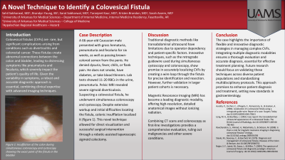
Case Description/Methods: A 66-year-old Caucasian male presented with gross hematuria, pneumaturia and fecaluria for six weeks, as well as passing brown-colored semen from the penis. He denied dysuria, fever, chills, or flank pain. He does not smoke, have diabetes, or take blood thinners. Lab tests showed 11-20 RBCs in the urine, pneumaturia. Pelvic MRI revealed severe sigmoid diverticulosis. Suspecting a colovesical fistula, he underwent simultaneous colonoscopy and cystoscopy. Despite extensive workup and initial difficulties locating the fistula, colonic insufflation localized it (figure 1). This novel technique allowed for direct visualization and successful surgical intervention through a robotic-assisted laparoscopic sigmoid colectomy.
Discussion: Traditional diagnostic methods like transabdominal ultrasound have limitations due to operator dependency and patient-specific factors. Innovative techniques, such as the retrograde guidewire used during simultaneous cystoscopy and colonoscopy, show promise in accurately localizing CVFs by creating a wire loop through the fistula for precise identification and resection. However, further validation in larger patient cohorts is necessary.
Magnetic Resonance Imaging (MRI) has become a leading diagnostic modality, offering high-resolution, detailed anatomical images without ionizing radiation.
Combining CT scans and colonoscopy as first-line investigations provides a comprehensive evaluation, ruling out malignancies and other severe conditions.
The case highlights the importance of flexible and innovative diagnostic strategies in managing complex CVFs. Integrating multiple diagnostic modalities ensures a thorough evaluation and accurate diagnosis, essential for effective treatment planning. Future research should focus on validating these techniques across diverse patient populations and standardizing multidisciplinary protocols. This approach promises to enhance patient diagnosis and treatment, setting new standards in gastroenterology.

Disclosures:
Sahil Sabharwal, MD1, Brandyn Young, BS1, Sarat Sabharwal, MD2, Taranjeet Kaur, MD2, Kristen Brandon, MD1, Sarah Assem, MD1. P3762 - A Novel Technique to Identify a Colovesical Fistula, ACG 2024 Annual Scientific Meeting Abstracts. Philadelphia, PA: American College of Gastroenterology.
1University of Arkansas for Medical Sciences, Fayetteville, AR; 2University of Kentucky College of Medicine, Hazard, KY
Introduction: Colovesical fistulas (CVFs) are rare, but significant complications arising from conditions such as diverticulitis and colorectal cancer. These fistulas create abnormal connections between the colon and bladder, leading to distressing symptoms like pneumaturia and fecaluria, which severely impact the patient's quality of life. Given the variability in symptoms, a robust and accurate diagnostic approach is essential, combining clinical expertise with advanced imaging techniques.
Case Description/Methods: A 66-year-old Caucasian male presented with gross hematuria, pneumaturia and fecaluria for six weeks, as well as passing brown-colored semen from the penis. He denied dysuria, fever, chills, or flank pain. He does not smoke, have diabetes, or take blood thinners. Lab tests showed 11-20 RBCs in the urine, pneumaturia. Pelvic MRI revealed severe sigmoid diverticulosis. Suspecting a colovesical fistula, he underwent simultaneous colonoscopy and cystoscopy. Despite extensive workup and initial difficulties locating the fistula, colonic insufflation localized it (figure 1). This novel technique allowed for direct visualization and successful surgical intervention through a robotic-assisted laparoscopic sigmoid colectomy.
Discussion: Traditional diagnostic methods like transabdominal ultrasound have limitations due to operator dependency and patient-specific factors. Innovative techniques, such as the retrograde guidewire used during simultaneous cystoscopy and colonoscopy, show promise in accurately localizing CVFs by creating a wire loop through the fistula for precise identification and resection. However, further validation in larger patient cohorts is necessary.
Magnetic Resonance Imaging (MRI) has become a leading diagnostic modality, offering high-resolution, detailed anatomical images without ionizing radiation.
Combining CT scans and colonoscopy as first-line investigations provides a comprehensive evaluation, ruling out malignancies and other severe conditions.
The case highlights the importance of flexible and innovative diagnostic strategies in managing complex CVFs. Integrating multiple diagnostic modalities ensures a thorough evaluation and accurate diagnosis, essential for effective treatment planning. Future research should focus on validating these techniques across diverse patient populations and standardizing multidisciplinary protocols. This approach promises to enhance patient diagnosis and treatment, setting new standards in gastroenterology.

Figure: Insufflation of the colon during simultaneous colonoscopy and cystoscopy showing the exact point of the fistula in the bladder.
Disclosures:
Sahil Sabharwal indicated no relevant financial relationships.
Brandyn Young indicated no relevant financial relationships.
Sarat Sabharwal indicated no relevant financial relationships.
Taranjeet Kaur indicated no relevant financial relationships.
Kristen Brandon indicated no relevant financial relationships.
Sarah Assem indicated no relevant financial relationships.
Sahil Sabharwal, MD1, Brandyn Young, BS1, Sarat Sabharwal, MD2, Taranjeet Kaur, MD2, Kristen Brandon, MD1, Sarah Assem, MD1. P3762 - A Novel Technique to Identify a Colovesical Fistula, ACG 2024 Annual Scientific Meeting Abstracts. Philadelphia, PA: American College of Gastroenterology.
