Tuesday Poster Session
Category: Colon
P3766 - Unmasking the Silent Invader: A Rare Case of Metastatic Appendiceal Goblet Cell Adenocarcinoma
Tuesday, October 29, 2024
10:30 AM - 4:00 PM ET
Location: Exhibit Hall E

Has Audio

Puja Sasankan, BS
George Washington University School of Medicine and Health Sciences
Washington, DC
Presenting Author(s)
Puja Sasankan, BS1, Priya Sasankan, MD, MBA2, Ibrahim Nassour, MD2, David Saulino, DO2, Ruby Deveras, MD3, Aleksey Novikov, MD2
1George Washington University School of Medicine and Health Sciences, Washington, DC; 2University of Florida College of Medicine, Gainesville, FL; 3Halifax Health, Daytona Beach, FL
Introduction: Goblet cell adenocarcinoma (GCA), a rare and aggressive tumor often originating from the appendix. We report on a 74-year-old female patient initially thought to have metastatic colon adenocarcinoma, later identified as having GCA with extensive lymph node involvement.
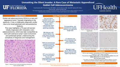
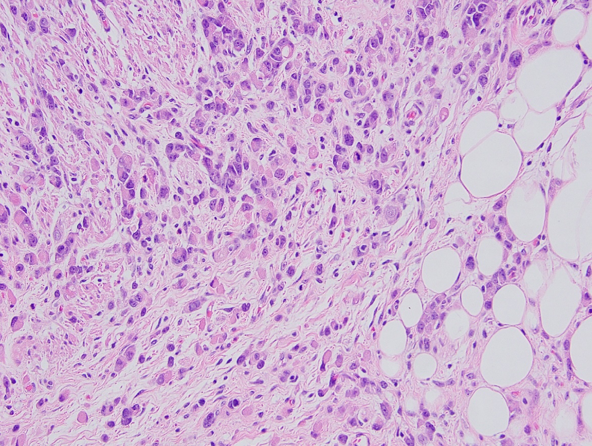
Case Description/Methods: A 74-year-old woman with a Roux-en-Y gastric bypass presented with months of diarrhea and weight loss. A year before symptom onset, a colonoscopy revealed one polyp, which was removed. One year later, she developed diarrhea with foul-smelling stools, leading to multiple office visits. An MR abdomen showed severe dilation of a small bowel loop with a small volume of free fluid and a CT abdomen and pelvis indicated mild colon distention without obstruction. Further investigation with an EGD and colonoscopy revealed ulcerations with skip lesions and severe angulated diverticulosis. Biopsies noted poorly differentiated adenocarcinoma with signet ring features. A PET scan indicated a tumor in the splenic flexure and hypermetabolic activity in the right lower quadrant of the small bowel. She underwent exploratory laparotomy, total abdominal colectomy, end ileostomy, and intraoperative colonoscopy. Pathology initially confirmed poorly differentiated invasive adenocarcinoma with metastatic involvement of all examined lymph nodes. Further evaluation with immunohistochemical staining indicated goblet cell adenocarcinoma, with positive markers for chromogranin, synaptophysin, CK7, CK20, and CDX2 (Image 1). Following surgery, she was seen for evaluation. Since she had not received chemotherapy, recommendation was for medical oncology follow-up, three months of chemotherapy, and re-evaluation for HIPEC.
Discussion: GCA is a rare neoplasm primarily found in the appendix, featuring both glandular and neuroendocrine characteristics, complicating its classification and diagnosis. In this case, initial diagnosis of metastatic colon adenocarcinoma was revised upon identifying signet ring – like cells and neuroendocrine markers. Identifying the tumor origin was challenging given the extensive intestinal involvement, however, the presence of goblet cells and neuroendocrine features suggested appendiceal origin. The lymph node involvement and peritoneal spread underscore the aggressive nature of this malignancy. Treatment strategies for GCA include cytoreductive surgery and HIPEC, followed by chemotherapy. This case reflects the need for comprehensive histopathological evaluation in advanced gastrointestinal malignancies.

Disclosures:
Puja Sasankan, BS1, Priya Sasankan, MD, MBA2, Ibrahim Nassour, MD2, David Saulino, DO2, Ruby Deveras, MD3, Aleksey Novikov, MD2. P3766 - Unmasking the Silent Invader: A Rare Case of Metastatic Appendiceal Goblet Cell Adenocarcinoma, ACG 2024 Annual Scientific Meeting Abstracts. Philadelphia, PA: American College of Gastroenterology.
1George Washington University School of Medicine and Health Sciences, Washington, DC; 2University of Florida College of Medicine, Gainesville, FL; 3Halifax Health, Daytona Beach, FL
Introduction: Goblet cell adenocarcinoma (GCA), a rare and aggressive tumor often originating from the appendix. We report on a 74-year-old female patient initially thought to have metastatic colon adenocarcinoma, later identified as having GCA with extensive lymph node involvement.
Case Description/Methods: A 74-year-old woman with a Roux-en-Y gastric bypass presented with months of diarrhea and weight loss. A year before symptom onset, a colonoscopy revealed one polyp, which was removed. One year later, she developed diarrhea with foul-smelling stools, leading to multiple office visits. An MR abdomen showed severe dilation of a small bowel loop with a small volume of free fluid and a CT abdomen and pelvis indicated mild colon distention without obstruction. Further investigation with an EGD and colonoscopy revealed ulcerations with skip lesions and severe angulated diverticulosis. Biopsies noted poorly differentiated adenocarcinoma with signet ring features. A PET scan indicated a tumor in the splenic flexure and hypermetabolic activity in the right lower quadrant of the small bowel. She underwent exploratory laparotomy, total abdominal colectomy, end ileostomy, and intraoperative colonoscopy. Pathology initially confirmed poorly differentiated invasive adenocarcinoma with metastatic involvement of all examined lymph nodes. Further evaluation with immunohistochemical staining indicated goblet cell adenocarcinoma, with positive markers for chromogranin, synaptophysin, CK7, CK20, and CDX2 (Image 1). Following surgery, she was seen for evaluation. Since she had not received chemotherapy, recommendation was for medical oncology follow-up, three months of chemotherapy, and re-evaluation for HIPEC.
Discussion: GCA is a rare neoplasm primarily found in the appendix, featuring both glandular and neuroendocrine characteristics, complicating its classification and diagnosis. In this case, initial diagnosis of metastatic colon adenocarcinoma was revised upon identifying signet ring – like cells and neuroendocrine markers. Identifying the tumor origin was challenging given the extensive intestinal involvement, however, the presence of goblet cells and neuroendocrine features suggested appendiceal origin. The lymph node involvement and peritoneal spread underscore the aggressive nature of this malignancy. Treatment strategies for GCA include cytoreductive surgery and HIPEC, followed by chemotherapy. This case reflects the need for comprehensive histopathological evaluation in advanced gastrointestinal malignancies.

Figure: Discohesive tumor cells representing goblet cell adenocarcinoma
Disclosures:
Puja Sasankan indicated no relevant financial relationships.
Priya Sasankan indicated no relevant financial relationships.
Ibrahim Nassour indicated no relevant financial relationships.
David Saulino indicated no relevant financial relationships.
Ruby Deveras indicated no relevant financial relationships.
Aleksey Novikov indicated no relevant financial relationships.
Puja Sasankan, BS1, Priya Sasankan, MD, MBA2, Ibrahim Nassour, MD2, David Saulino, DO2, Ruby Deveras, MD3, Aleksey Novikov, MD2. P3766 - Unmasking the Silent Invader: A Rare Case of Metastatic Appendiceal Goblet Cell Adenocarcinoma, ACG 2024 Annual Scientific Meeting Abstracts. Philadelphia, PA: American College of Gastroenterology.
