Tuesday Poster Session
Category: Colon
P3790 - A Rare Case of Inflammatory Fibroid Polyp Mimicking Malignancy on Colonoscopy
Tuesday, October 29, 2024
10:30 AM - 4:00 PM ET
Location: Exhibit Hall E

Has Audio

Muhammad YN Chaudhary, MBChB
Indiana University Southwest
Evansville, IN
Presenting Author(s)
Muhammad YN. Chaudhary, MBChB1, Muhammad Ismail, MD2, Oluwagbenga Serrano, MD, FACG3
1Indiana University Southwest, Evansville, IN; 2Indiana University Southwest, Cedar Rapids, IA; 3Good Samaritan Hospital, Vincennes, IN
Introduction: Inflammatory fibroid polyps (IFPs) are rare, benign tumors originating from the submucosa of the gastrointestinal (GI) tract. Though most commonly found in the stomach, IFPs can occur throughout the GI tract, including the colon. Due to their rarity and variable presentation, IFPs can be difficult to diagnose, often mimicking malignant lesions on endoscopic evaluation. This case report presents a novel instance of an IFP in the srectum, initially suspected to be malignant, and underscores the importance of histopathological confirmation to avoid misdiagnosis and unnecessary intervention.
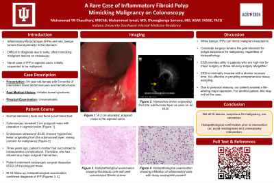
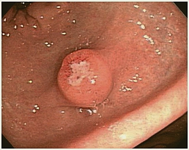
Case Description/Methods: A 54-year-old female with a history of irritable bowel syndrome presented with a six-month history of intermittent lower abdominal pain and hematochezia. Physical examination was unremarkable. All laboratory tests and fecal occult blood testing were within normal limits. Given her symptoms, a colonoscopy was performed, revealing a 3 cm polypoid mass with ulceration in the rectum (Image 1).
Endoscopic ultrasound (EUS) demonstrated a hypoechoic lesion originating from the submucosal layer, raising concern for a potentially malignant lesion. The patient had previously refused major surgery as her mother succumbed to intraoperative complications three years ago. As such, our patient underwent an endoscopic surgical dissection (ESD) of the polypoid mass. Histopathological examination revealed a submucosal proliferation of spindle cells with a prominent inflammatory infiltrate, consistent with the diagnosis of an IFP.
Discussion: IFPs are characterized histologically by a proliferation of fibroblasts, myofibroblasts, and inflammatory cells. While benign, IFPs can present with endoscopic appearances that mimic malignant neoplasms. Although colorectal surgery remains the gold-standard for polyps suspicious for malignancy, ESD provides utility in patients who are otherwise high-risk for major surgery or those patients refusing surgery altogether. This was the case with our patient.
Compared to traditional surgical resection, ESD offers the advantage of being minimally invasive with a shorter recovery time. It is still effective in providing comprehensive tissue sampling necessary for accurate histopathological assessment.
The patient’s lesion, with its ulcerated surface and submucosal origin, closely resembled malignancy. Awareness of IFPs and their diverse presentations is crucial in avoiding misdiagnosis of complex GI polyps and unnecessary invasive procedures.

Disclosures:
Muhammad YN. Chaudhary, MBChB1, Muhammad Ismail, MD2, Oluwagbenga Serrano, MD, FACG3. P3790 - A Rare Case of Inflammatory Fibroid Polyp Mimicking Malignancy on Colonoscopy, ACG 2024 Annual Scientific Meeting Abstracts. Philadelphia, PA: American College of Gastroenterology.
1Indiana University Southwest, Evansville, IN; 2Indiana University Southwest, Cedar Rapids, IA; 3Good Samaritan Hospital, Vincennes, IN
Introduction: Inflammatory fibroid polyps (IFPs) are rare, benign tumors originating from the submucosa of the gastrointestinal (GI) tract. Though most commonly found in the stomach, IFPs can occur throughout the GI tract, including the colon. Due to their rarity and variable presentation, IFPs can be difficult to diagnose, often mimicking malignant lesions on endoscopic evaluation. This case report presents a novel instance of an IFP in the srectum, initially suspected to be malignant, and underscores the importance of histopathological confirmation to avoid misdiagnosis and unnecessary intervention.
Case Description/Methods: A 54-year-old female with a history of irritable bowel syndrome presented with a six-month history of intermittent lower abdominal pain and hematochezia. Physical examination was unremarkable. All laboratory tests and fecal occult blood testing were within normal limits. Given her symptoms, a colonoscopy was performed, revealing a 3 cm polypoid mass with ulceration in the rectum (Image 1).
Endoscopic ultrasound (EUS) demonstrated a hypoechoic lesion originating from the submucosal layer, raising concern for a potentially malignant lesion. The patient had previously refused major surgery as her mother succumbed to intraoperative complications three years ago. As such, our patient underwent an endoscopic surgical dissection (ESD) of the polypoid mass. Histopathological examination revealed a submucosal proliferation of spindle cells with a prominent inflammatory infiltrate, consistent with the diagnosis of an IFP.
Discussion: IFPs are characterized histologically by a proliferation of fibroblasts, myofibroblasts, and inflammatory cells. While benign, IFPs can present with endoscopic appearances that mimic malignant neoplasms. Although colorectal surgery remains the gold-standard for polyps suspicious for malignancy, ESD provides utility in patients who are otherwise high-risk for major surgery or those patients refusing surgery altogether. This was the case with our patient.
Compared to traditional surgical resection, ESD offers the advantage of being minimally invasive with a shorter recovery time. It is still effective in providing comprehensive tissue sampling necessary for accurate histopathological assessment.
The patient’s lesion, with its ulcerated surface and submucosal origin, closely resembled malignancy. Awareness of IFPs and their diverse presentations is crucial in avoiding misdiagnosis of complex GI polyps and unnecessary invasive procedures.

Figure: Image 1: Ulcerated polypoid mass in the rectum.
Disclosures:
Muhammad Chaudhary indicated no relevant financial relationships.
Muhammad Ismail indicated no relevant financial relationships.
Oluwagbenga Serrano: MERCK – Stock-publicly held company(excluding mutual/index funds).
Muhammad YN. Chaudhary, MBChB1, Muhammad Ismail, MD2, Oluwagbenga Serrano, MD, FACG3. P3790 - A Rare Case of Inflammatory Fibroid Polyp Mimicking Malignancy on Colonoscopy, ACG 2024 Annual Scientific Meeting Abstracts. Philadelphia, PA: American College of Gastroenterology.
