Tuesday Poster Session
Category: Colon
P3804 - Diagnosing Sigmoid Endometriosis by EUS-FNB
Tuesday, October 29, 2024
10:30 AM - 4:00 PM ET
Location: Exhibit Hall E

Has Audio

Matthew M. Eganhouse, MD
Rush University Medical Center
Chicago, IL
Presenting Author(s)
Matthew M. Eganhouse, MD, Agnieska Maniak, MD, Isabel Hart, MD, Neal A. Mehta, MD, Christopher Chapman, MD, Ajaypal Singh, MD, Irving Waxman, MD
Rush University Medical Center, Chicago, IL
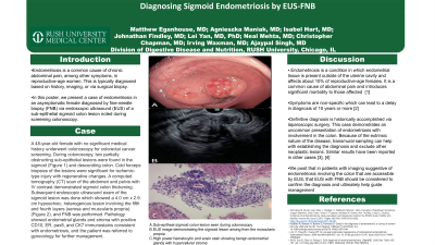
Introduction: Endometriosis is a common cause of chronic abdominal pain, among other symptoms, in reproductive-age women. Typically, this condition is either diagnosed clinically based on history, exam, and imaging, or via surgical visualization or biopsy. In this report, we describe a case of endometriosis in an asymptomatic female diagnosed by fine-needle biopsy (FNB) via endoscopic ultrasound (EUS) of a sigmoid colon sub-epithelial lesion noted incidentally during screening colonoscopy.
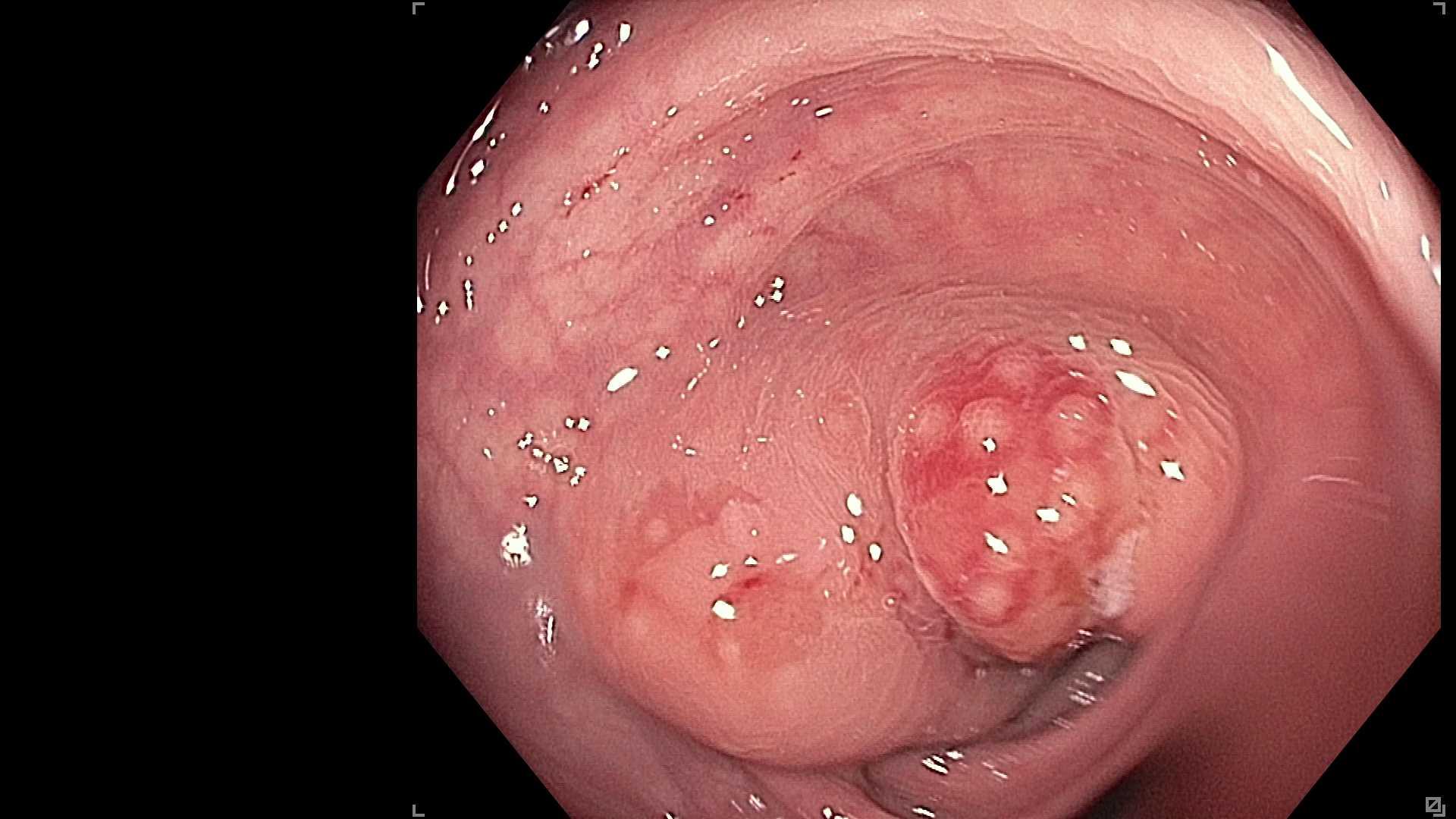
Case Description/Methods: A 48 year old female with no significant medical history underwent colonoscopy for colorectal cancer screening. During colonoscopy, two partially obstructing sub-epithelial lesions were found in the sigmoid (Figure 1) and descending colon. Cold forceps biopsies of the lesions were significant for ischemic-type injury with regenerative changes. A computed tomography (CT) scan of the abdomen and pelvis with IV contrast demonstrated sigmoid colon thickening. Subsequent endoscopic ultrasound exam of the sigmoid lesion was done which showed a 4.0 cm x 2.9. cm hypoechoic, heterogenous lesion involving the fifth and fourth layers (serosa and muscularis propria), and FNB was performed. Pathology showed endometrial glands and stroma with positive CD10, ER, pax8, and CK7 immunostains consistent with endometriosis, and the patient was referred to gynecology for further management.
Discussion: Endometriosis is a condition in which endometrial tissue is present outside of the uterine cavity. This disease affects about 10% of reproductive-age females and is a common cause of chronic abdominal pain, among other symptoms, and causes significant morbidity. Symptoms are non-specific which can lead to a delay in clinical diagnosis of 10 years or more. Definitive diagnosis is historically accomplished via laparoscopic surgery, which is invasive and can be frightening for patients. This case demonstrates an uncommon presentation of endometriosis with involvement of the colonic wall. Due to the extrinsic nature of the lesion, endoscopic biopsies are usually diagnostic and EUS guided transmural sampling can help with establishing diagnosis and exclude other neoplastic lesions. There have been other case reports with similar results. Therefore, in patients with imaging suggestive of endometriosis involving the colon, particularly in areas reachable by EUS, EUS with FNB should be considered to confirm the diagnosis and ultimately help guide management.

Disclosures:
Matthew M. Eganhouse, MD, Agnieska Maniak, MD, Isabel Hart, MD, Neal A. Mehta, MD, Christopher Chapman, MD, Ajaypal Singh, MD, Irving Waxman, MD. P3804 - Diagnosing Sigmoid Endometriosis by EUS-FNB, ACG 2024 Annual Scientific Meeting Abstracts. Philadelphia, PA: American College of Gastroenterology.
Rush University Medical Center, Chicago, IL
Introduction: Endometriosis is a common cause of chronic abdominal pain, among other symptoms, in reproductive-age women. Typically, this condition is either diagnosed clinically based on history, exam, and imaging, or via surgical visualization or biopsy. In this report, we describe a case of endometriosis in an asymptomatic female diagnosed by fine-needle biopsy (FNB) via endoscopic ultrasound (EUS) of a sigmoid colon sub-epithelial lesion noted incidentally during screening colonoscopy.
Case Description/Methods: A 48 year old female with no significant medical history underwent colonoscopy for colorectal cancer screening. During colonoscopy, two partially obstructing sub-epithelial lesions were found in the sigmoid (Figure 1) and descending colon. Cold forceps biopsies of the lesions were significant for ischemic-type injury with regenerative changes. A computed tomography (CT) scan of the abdomen and pelvis with IV contrast demonstrated sigmoid colon thickening. Subsequent endoscopic ultrasound exam of the sigmoid lesion was done which showed a 4.0 cm x 2.9. cm hypoechoic, heterogenous lesion involving the fifth and fourth layers (serosa and muscularis propria), and FNB was performed. Pathology showed endometrial glands and stroma with positive CD10, ER, pax8, and CK7 immunostains consistent with endometriosis, and the patient was referred to gynecology for further management.
Discussion: Endometriosis is a condition in which endometrial tissue is present outside of the uterine cavity. This disease affects about 10% of reproductive-age females and is a common cause of chronic abdominal pain, among other symptoms, and causes significant morbidity. Symptoms are non-specific which can lead to a delay in clinical diagnosis of 10 years or more. Definitive diagnosis is historically accomplished via laparoscopic surgery, which is invasive and can be frightening for patients. This case demonstrates an uncommon presentation of endometriosis with involvement of the colonic wall. Due to the extrinsic nature of the lesion, endoscopic biopsies are usually diagnostic and EUS guided transmural sampling can help with establishing diagnosis and exclude other neoplastic lesions. There have been other case reports with similar results. Therefore, in patients with imaging suggestive of endometriosis involving the colon, particularly in areas reachable by EUS, EUS with FNB should be considered to confirm the diagnosis and ultimately help guide management.

Figure: Sigmoid Colon Sub-Epithelial lesion
Disclosures:
Matthew Eganhouse indicated no relevant financial relationships.
Agnieska Maniak indicated no relevant financial relationships.
Isabel Hart indicated no relevant financial relationships.
Neal Mehta: Boston Scientific – Consultant. Castle Biosciences – Consultant. Conmed – Consultant. Medtronic – Consultant.
Christopher Chapman: Apollo Endosurgery – Consultant. Boston Scientific – Consultant. Olympus – Consultant.
Ajaypal Singh indicated no relevant financial relationships.
Irving Waxman indicated no relevant financial relationships.
Matthew M. Eganhouse, MD, Agnieska Maniak, MD, Isabel Hart, MD, Neal A. Mehta, MD, Christopher Chapman, MD, Ajaypal Singh, MD, Irving Waxman, MD. P3804 - Diagnosing Sigmoid Endometriosis by EUS-FNB, ACG 2024 Annual Scientific Meeting Abstracts. Philadelphia, PA: American College of Gastroenterology.
