Tuesday Poster Session
Category: Esophagus
P3917 - Topical Steroids in Lymphocytic Esophagitis: Worthy of the Prior Authorization?
Tuesday, October 29, 2024
10:30 AM - 4:00 PM ET
Location: Exhibit Hall E

Has Audio
- LL
Laura E. Lavette, MD
University of Virginia Medical Center
Charlottesville, VA
Presenting Author(s)
Laura E. Lavette, MD, Sahil Khanna, MD, Chris Young, MD, Calvin Geng, MD, Amanda Gibbs, MD, Anne Mills, MD, Edward Stelow, MD, Andrew Copland, MD
University of Virginia Medical Center, Charlottesville, VA
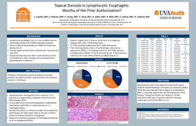
Introduction: Lymphocytic esophagitis (LE) is a rare condition with an estimated incidence of 1/1000 esophageal biopsies. There is little existing literature on effective treatment options for LE. Previous patients have shown symptomatic improvement with PPIs. Topical steroids have also been recommended, as they are beneficial in other autoimmune and lymphocytic gastrointestinal conditions.1-3 Through a retrospective review of 17 pediatric and adult patients, we aimed to better understand the role of topical steroids in treating LE.
Methods: We performed a retrospective chart review of 17 LE patients at a single tertiary academic center from June 2012-Oct 2022. LE was defined as increased peripapillary intraepithelial lymphocytes with little or no granulocytes on ≥1 esophageal biopsy; we did not enforce a minimum number of intraepithelial lymphocytes per high power field, as this has not been shown to improve specificity of diagnosis.4 Response to therapy was based on histological resolution of LE on subsequent EGD(s).
Results: Patients ranged from ages 3-74 at time of LE diagnosis, with 7 (41%) being male. All 17 patients were on a PPI prior to diagnosis. 12 (71%) patients were treated with budesonide and 5 (29%) with fluticasone. From the budesonide cohort, 4 (33%) had histologic remission on subsequent EGD(s), 5 (42%) showed persistent LE, and 3 (25%) did not have histologic data available. In the fluticasone group, 2 (40%) had histologic remission, 2 (40%) did not, and 1 (20%) was without follow-up EGD(s).
Discussion: Approximately half of the patients treated with topical steroids showed histologic remission on subsequent EGD(s). While this may represent some degree of a therapeutic effect, it may also support the self-limiting nature of the disease. Prospective studies are needed to further characterize the role of steroids and their histologic, endoscopic, and clinical effects on LE.
1. Pasricha S, et al. Lymphocytic Esophagitis: An Emerging Clinicopathologic Disease Associated with Dysphagia. Dig Dis Sci. 2016 Oct;61(10):2935–41.
2. Kasirye Y, et al. Lymphocytic Esophagitis Presenting as Chronic Dysphagia. Clin Med Res. 2012 May;10(2):83–4.
3. Romana B, et al. Lymphocytic Esophagitis: Steroids Make a Difference. Amer Jour Gastro. 2016 Oct;111():S776-S777.
4. Haque S, Genta RM. Lymphocytic oesophagitis: clinicopathological aspects of an emerging condition. Gut. 2012 Aug;61(8):1108–14.

Note: The table for this abstract can be viewed in the ePoster Gallery section of the ACG 2024 ePoster Site or in The American Journal of Gastroenterology's abstract supplement issue, both of which will be available starting October 27, 2024.
Disclosures:
Laura E. Lavette, MD, Sahil Khanna, MD, Chris Young, MD, Calvin Geng, MD, Amanda Gibbs, MD, Anne Mills, MD, Edward Stelow, MD, Andrew Copland, MD. P3917 - Topical Steroids in Lymphocytic Esophagitis: Worthy of the Prior Authorization?, ACG 2024 Annual Scientific Meeting Abstracts. Philadelphia, PA: American College of Gastroenterology.
University of Virginia Medical Center, Charlottesville, VA
Introduction: Lymphocytic esophagitis (LE) is a rare condition with an estimated incidence of 1/1000 esophageal biopsies. There is little existing literature on effective treatment options for LE. Previous patients have shown symptomatic improvement with PPIs. Topical steroids have also been recommended, as they are beneficial in other autoimmune and lymphocytic gastrointestinal conditions.1-3 Through a retrospective review of 17 pediatric and adult patients, we aimed to better understand the role of topical steroids in treating LE.
Methods: We performed a retrospective chart review of 17 LE patients at a single tertiary academic center from June 2012-Oct 2022. LE was defined as increased peripapillary intraepithelial lymphocytes with little or no granulocytes on ≥1 esophageal biopsy; we did not enforce a minimum number of intraepithelial lymphocytes per high power field, as this has not been shown to improve specificity of diagnosis.4 Response to therapy was based on histological resolution of LE on subsequent EGD(s).
Results: Patients ranged from ages 3-74 at time of LE diagnosis, with 7 (41%) being male. All 17 patients were on a PPI prior to diagnosis. 12 (71%) patients were treated with budesonide and 5 (29%) with fluticasone. From the budesonide cohort, 4 (33%) had histologic remission on subsequent EGD(s), 5 (42%) showed persistent LE, and 3 (25%) did not have histologic data available. In the fluticasone group, 2 (40%) had histologic remission, 2 (40%) did not, and 1 (20%) was without follow-up EGD(s).
Discussion: Approximately half of the patients treated with topical steroids showed histologic remission on subsequent EGD(s). While this may represent some degree of a therapeutic effect, it may also support the self-limiting nature of the disease. Prospective studies are needed to further characterize the role of steroids and their histologic, endoscopic, and clinical effects on LE.
1. Pasricha S, et al. Lymphocytic Esophagitis: An Emerging Clinicopathologic Disease Associated with Dysphagia. Dig Dis Sci. 2016 Oct;61(10):2935–41.
2. Kasirye Y, et al. Lymphocytic Esophagitis Presenting as Chronic Dysphagia. Clin Med Res. 2012 May;10(2):83–4.
3. Romana B, et al. Lymphocytic Esophagitis: Steroids Make a Difference. Amer Jour Gastro. 2016 Oct;111():S776-S777.
4. Haque S, Genta RM. Lymphocytic oesophagitis: clinicopathological aspects of an emerging condition. Gut. 2012 Aug;61(8):1108–14.

Figure: Figure 1. Histologic sections show stratified squamous epithelium with numerous intraepithelial lymphocytes and intraepithelial edema (spongiosis) with occasional dyskeratotic cells.
Note: The table for this abstract can be viewed in the ePoster Gallery section of the ACG 2024 ePoster Site or in The American Journal of Gastroenterology's abstract supplement issue, both of which will be available starting October 27, 2024.
Disclosures:
Laura Lavette indicated no relevant financial relationships.
Sahil Khanna indicated no relevant financial relationships.
Chris Young indicated no relevant financial relationships.
Calvin Geng indicated no relevant financial relationships.
Amanda Gibbs indicated no relevant financial relationships.
Anne Mills indicated no relevant financial relationships.
Edward Stelow indicated no relevant financial relationships.
Andrew Copland indicated no relevant financial relationships.
Laura E. Lavette, MD, Sahil Khanna, MD, Chris Young, MD, Calvin Geng, MD, Amanda Gibbs, MD, Anne Mills, MD, Edward Stelow, MD, Andrew Copland, MD. P3917 - Topical Steroids in Lymphocytic Esophagitis: Worthy of the Prior Authorization?, ACG 2024 Annual Scientific Meeting Abstracts. Philadelphia, PA: American College of Gastroenterology.
