Tuesday Poster Session
Category: Esophagus
P3954 - A Case of Dysphagia due to an Aberrant Right Subclavian Artery: Dysphagia Lusoria
Tuesday, October 29, 2024
10:30 AM - 4:00 PM ET
Location: Exhibit Hall E


Jonathan Rozenberg, DO, MPH
Carilion Clinic
Roanoke, VA
Presenting Author(s)
Jonathan Rozenberg, DO, MPH1, Adil Mir, MD1, Paul Yeaton, MD2, Joseph Birkman, DO3
1Carilion Clinic, Roanoke, VA; 2Carilion Clinic Virginia Tech, Roanoke, VA; 3Virginia Tech Carilion School of Medicine, Roanoke, VA
Introduction: Extrinsic esophageal compression by surrounding vasculature resulting in dysphagia is an uncommon occurrence. The most common type of aortic arch embryologic defect is an aberrant right subclavian artery (ARSA) which runs retroesophageal. ARSAs are typically found incidentally and 60-80% of those with this anomaly are asymptomatic. Dysphagia lusoria refers to the phenomena of dysphagia secondary to esophageal compression by the trachea anteriorly and an ARSA posteriorly, which is often seen on barium esophagram. We present a rare case of dysphagia in the setting of a retroesophageal ARSA with corroborating radiographic evidence.
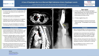
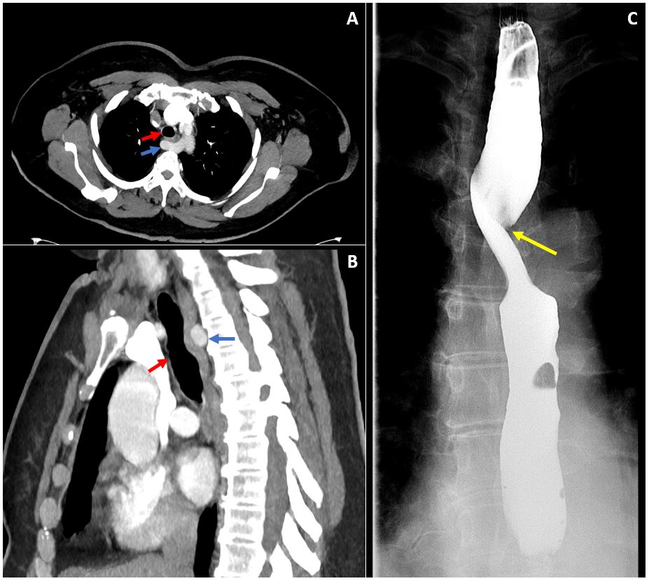
Case Description/Methods: A 65-year-old female presented for evaluation of subacute dysphagia after an ARSA was identified on MRI Chest done for evaluation of right sternoclavicular joint swelling and on follow up CTA Chest (Figure 1). Past medical history was significant for chronic GERD. She was evaluated for dysphagia and globus sensation 5 years prior via a barium esophagram that demonstrated significant reflux to the level of the thoracic inlet without indentation or evidence of esophageal obstruction. EGD at that time found erosive gastropathy in the gastric antrum. Conservative measures (e.g., small frequent meals) and acid suppression therapy with esomeprazole 20 mg was recommended. Repeat barium esophagram saw right lateral displacement and narrowing of the esophagus adjacent to the aortic knob with "bayonet sign" appearance consistent with known ARSA posterior to the esophagus (Figure 1). Patient’s symptoms and radiographic evidence consistent with a diagnosis of dysphagia lusoria.
Discussion: When evaluating dysphagia, its underlying mechanism influences diagnostic workup and management greatly. In dysphagia lusoria, both physical exam and EGD are often largely unremarkable with multimodal imaging involving a barium esophagram alongside CT or MRI imaging being the highest yield studies necessary for diagnosis. Notably, endoscopic ultrasound does have utility in this scenario as it can aid in verifying a vascular component if esophageal compression is appreciated on endoscopy. Our case highlights the need to maintain a broad differential in dysphagia work-up.

Disclosures:
Jonathan Rozenberg, DO, MPH1, Adil Mir, MD1, Paul Yeaton, MD2, Joseph Birkman, DO3. P3954 - A Case of Dysphagia due to an Aberrant Right Subclavian Artery: Dysphagia Lusoria, ACG 2024 Annual Scientific Meeting Abstracts. Philadelphia, PA: American College of Gastroenterology.
1Carilion Clinic, Roanoke, VA; 2Carilion Clinic Virginia Tech, Roanoke, VA; 3Virginia Tech Carilion School of Medicine, Roanoke, VA
Introduction: Extrinsic esophageal compression by surrounding vasculature resulting in dysphagia is an uncommon occurrence. The most common type of aortic arch embryologic defect is an aberrant right subclavian artery (ARSA) which runs retroesophageal. ARSAs are typically found incidentally and 60-80% of those with this anomaly are asymptomatic. Dysphagia lusoria refers to the phenomena of dysphagia secondary to esophageal compression by the trachea anteriorly and an ARSA posteriorly, which is often seen on barium esophagram. We present a rare case of dysphagia in the setting of a retroesophageal ARSA with corroborating radiographic evidence.
Case Description/Methods: A 65-year-old female presented for evaluation of subacute dysphagia after an ARSA was identified on MRI Chest done for evaluation of right sternoclavicular joint swelling and on follow up CTA Chest (Figure 1). Past medical history was significant for chronic GERD. She was evaluated for dysphagia and globus sensation 5 years prior via a barium esophagram that demonstrated significant reflux to the level of the thoracic inlet without indentation or evidence of esophageal obstruction. EGD at that time found erosive gastropathy in the gastric antrum. Conservative measures (e.g., small frequent meals) and acid suppression therapy with esomeprazole 20 mg was recommended. Repeat barium esophagram saw right lateral displacement and narrowing of the esophagus adjacent to the aortic knob with "bayonet sign" appearance consistent with known ARSA posterior to the esophagus (Figure 1). Patient’s symptoms and radiographic evidence consistent with a diagnosis of dysphagia lusoria.
Discussion: When evaluating dysphagia, its underlying mechanism influences diagnostic workup and management greatly. In dysphagia lusoria, both physical exam and EGD are often largely unremarkable with multimodal imaging involving a barium esophagram alongside CT or MRI imaging being the highest yield studies necessary for diagnosis. Notably, endoscopic ultrasound does have utility in this scenario as it can aid in verifying a vascular component if esophageal compression is appreciated on endoscopy. Our case highlights the need to maintain a broad differential in dysphagia work-up.

Figure: Figure 1: Panel A) CT Chest, Axial View, and B) Sagittal View showing esophageal flattening between the trachea anteriorly (red arrow) and the aberrant right subclavian artery posteriorly (blue arrow), C) Barium esophagram showing characteristic “bayonet sign” of Dysphagia Lusoria (yellow arrow)
Disclosures:
Jonathan Rozenberg indicated no relevant financial relationships.
Adil Mir indicated no relevant financial relationships.
Paul Yeaton indicated no relevant financial relationships.
Joseph Birkman indicated no relevant financial relationships.
Jonathan Rozenberg, DO, MPH1, Adil Mir, MD1, Paul Yeaton, MD2, Joseph Birkman, DO3. P3954 - A Case of Dysphagia due to an Aberrant Right Subclavian Artery: Dysphagia Lusoria, ACG 2024 Annual Scientific Meeting Abstracts. Philadelphia, PA: American College of Gastroenterology.
