Tuesday Poster Session
Category: Esophagus
P3961 - Unexpected Visitors: A Primary Esophageal Melanoma
Tuesday, October 29, 2024
10:30 AM - 4:00 PM ET
Location: Exhibit Hall E

Has Audio

Sanad Dawod, MD
Henry Ford Health
Detroit, MI
Presenting Author(s)
Sanad Dawod, MD1, Suhib Alhaj Ali, MD2, Noor Khalil, 1, Stephanie Betcher, MD3, Tingting Xiong, MD1, Keith Mullins, MD2
1Henry Ford Health, Detroit, MI; 2Henry Ford Hospital, Detroit, MI; 3Henry Ford Health, Royal Oak, MI
Introduction: Melanoma, a malignancy arising from melanocytes, is predominantly associated with the skin, yet its occurrence in extracutaneous sites such as the esophagus is rare, constituting less than 1% of esophageal malignancies. As of 2021, only 347 cases have been reported in the literature. Endoscopically, esophageal melanoma presents with varied appearances, from pigmented spots and patches to amelanotic masses, posing challenges in differentiation from other esophageal tumors. We present a case of primary esophageal melanoma in an 81-year old patient.
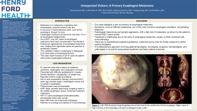
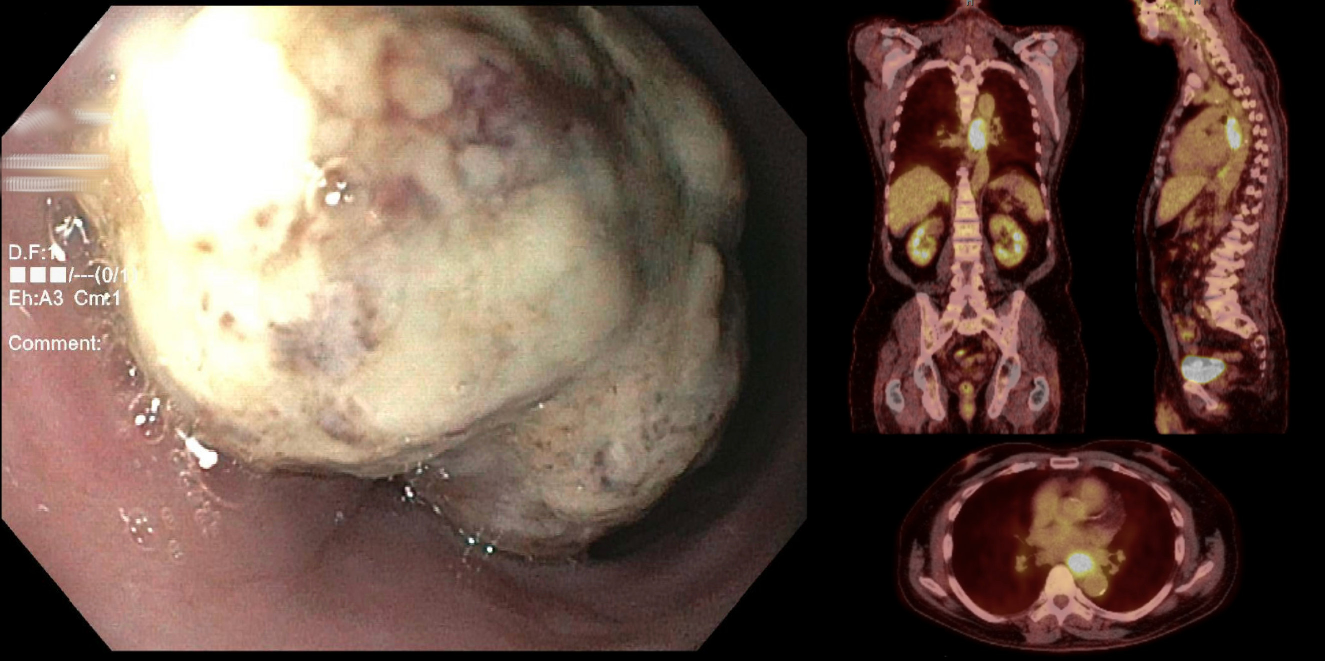
Case Description/Methods: Our patient is an 81-year-old male with history of metabolic syndrome, esophagitis, and cryptogenic cirrhosis. He presented to the office for evaluation of difficulty swallowing of 3 weeks duration. He denied heartburn, regurgitation, or weight loss. He endorsed preceding cough and fatigue for months prior as well. He was not on any acid suppression therapy. His last esophagogastroduodenoscopy (EGD) was 2 years prior, and was only remarkable for grade I esophageal varices, as well LA grade A esophagitis. His physical exam was unremarkable. He underwent an esophagogastroduodenoscopy (EGD) which showed a large fungating mass that was partially obstructing and partially circumferential, at the level of the middle esophagus. (Figure 1.) The mass was sampled, and was consistent with a malignant melanoma. He subsequently underwent full body imaging with PET-CT which was remarkable for paraesophageal lymphadenopathy. (Figure 2.) Dermatologic exam was normal. Brain MRI ruled out intracranial metastasis. The patient was referred to oncology and was initiated on immunotherapy.
Discussion: Our case highlights a rare instance of esophageal melanoma. Presenting symptoms, such as difficulty swallowing in our 81-year-old patient, often mimic more common esophageal conditions, emphasizing the diagnostic challenges. Esophageal melanomas tend to be aggressive, as evident by his normal EGD 2 years prior, with a high risk for metastasis. Therapeutic options for esophageal melanoma remain challenging due to its rarity, with surgery being a cornerstone, often complemented by immunotherapies. However, the absence of standardized treatment guidelines accentuates the need for ongoing research to establish optimal therapeutic strategies. Its rarity calls for a multidisciplinary approach involving gastroenterologists, oncologists, surgeons, dermatologists and pathologists in forming personalized treatment plans for optimal patient outcomes.

Disclosures:
Sanad Dawod, MD1, Suhib Alhaj Ali, MD2, Noor Khalil, 1, Stephanie Betcher, MD3, Tingting Xiong, MD1, Keith Mullins, MD2. P3961 - Unexpected Visitors: A Primary Esophageal Melanoma, ACG 2024 Annual Scientific Meeting Abstracts. Philadelphia, PA: American College of Gastroenterology.
1Henry Ford Health, Detroit, MI; 2Henry Ford Hospital, Detroit, MI; 3Henry Ford Health, Royal Oak, MI
Introduction: Melanoma, a malignancy arising from melanocytes, is predominantly associated with the skin, yet its occurrence in extracutaneous sites such as the esophagus is rare, constituting less than 1% of esophageal malignancies. As of 2021, only 347 cases have been reported in the literature. Endoscopically, esophageal melanoma presents with varied appearances, from pigmented spots and patches to amelanotic masses, posing challenges in differentiation from other esophageal tumors. We present a case of primary esophageal melanoma in an 81-year old patient.
Case Description/Methods: Our patient is an 81-year-old male with history of metabolic syndrome, esophagitis, and cryptogenic cirrhosis. He presented to the office for evaluation of difficulty swallowing of 3 weeks duration. He denied heartburn, regurgitation, or weight loss. He endorsed preceding cough and fatigue for months prior as well. He was not on any acid suppression therapy. His last esophagogastroduodenoscopy (EGD) was 2 years prior, and was only remarkable for grade I esophageal varices, as well LA grade A esophagitis. His physical exam was unremarkable. He underwent an esophagogastroduodenoscopy (EGD) which showed a large fungating mass that was partially obstructing and partially circumferential, at the level of the middle esophagus. (Figure 1.) The mass was sampled, and was consistent with a malignant melanoma. He subsequently underwent full body imaging with PET-CT which was remarkable for paraesophageal lymphadenopathy. (Figure 2.) Dermatologic exam was normal. Brain MRI ruled out intracranial metastasis. The patient was referred to oncology and was initiated on immunotherapy.
Discussion: Our case highlights a rare instance of esophageal melanoma. Presenting symptoms, such as difficulty swallowing in our 81-year-old patient, often mimic more common esophageal conditions, emphasizing the diagnostic challenges. Esophageal melanomas tend to be aggressive, as evident by his normal EGD 2 years prior, with a high risk for metastasis. Therapeutic options for esophageal melanoma remain challenging due to its rarity, with surgery being a cornerstone, often complemented by immunotherapies. However, the absence of standardized treatment guidelines accentuates the need for ongoing research to establish optimal therapeutic strategies. Its rarity calls for a multidisciplinary approach involving gastroenterologists, oncologists, surgeons, dermatologists and pathologists in forming personalized treatment plans for optimal patient outcomes.

Figure: Left: Esophagogastroduodenoscopy (EGD) showing a large fungating mass at the level of the middle third of the esophagus.
Right: PET-CT showing hyper-metabolic esophageal mass and paraesophageal lymphadenopathy.
Right: PET-CT showing hyper-metabolic esophageal mass and paraesophageal lymphadenopathy.
Disclosures:
Sanad Dawod indicated no relevant financial relationships.
Suhib Alhaj Ali indicated no relevant financial relationships.
Noor Khalil indicated no relevant financial relationships.
Stephanie Betcher indicated no relevant financial relationships.
Tingting Xiong indicated no relevant financial relationships.
Keith Mullins indicated no relevant financial relationships.
Sanad Dawod, MD1, Suhib Alhaj Ali, MD2, Noor Khalil, 1, Stephanie Betcher, MD3, Tingting Xiong, MD1, Keith Mullins, MD2. P3961 - Unexpected Visitors: A Primary Esophageal Melanoma, ACG 2024 Annual Scientific Meeting Abstracts. Philadelphia, PA: American College of Gastroenterology.

