Tuesday Poster Session
Category: Colon
P3770 - An Atypical Case of Abdominal Pain Secondary to Large Retroperitoneal Cyst
Tuesday, October 29, 2024
10:30 AM - 4:00 PM ET
Location: Exhibit Hall E

Has Audio

Haarika Korlipara, MD
New York-Presbyterian / Weill Cornell Medical Center
New York, NY
Presenting Author(s)
Haarika Korlipara, MD1, Douglas Scherr, MD2, Paul Fenyves, MD2
1New York-Presbyterian / Weill Cornell Medical Center, New York, NY; 2Weill Cornell Medicine, New York, NY
Introduction: Retroperitoneal (RP) cystic lesions are an uncommon and heterogeneous group of lesions that are derived from remnants of embryonal urogenital apparatus. They originate within the RP space, which is a complex region located behind the peritoneum, and this allows cysts to grow to a considerable size before becoming symptomatic. Here, we describe an atypical case of a symptomatic large RP cyst in a young woman.
Case Description/Methods: A 29-year-old woman with no significant past medical history presented to her PCP for two weeks of sharp left upper quadrant (LUQ) abdominal pain after a Pure Barre class. It was exacerbated by exertion and improved with rest. Physical exam was notable for tenderness to palpation above the left clavicle and left upper quadrant (LUQ) with no palpable abnormality, CVA tenderness, or splenomegaly. Labs were notable for WBC count 5.1, D-dimer < 0.19, and negative urine analysis. Her presentation was thought to be consistent with musculoskeletal pain and she was advised conservative management. Her pain didn’t improve, and she was ordered for an abdominal ultrasound which showed a LUQ cyst measuring 7.1cm and abutting the upper pole of the left kidney, spleen, and pancreatic tail. MRI abdomen with contrast showed a 7.7 x 6.0 x 6.5cm simple thin-walled cyst with no solid or nodular enhancement, likely reflecting a benign RP cyst. She was referred to urology who recommended a stability scan at 6 months and drainage if her symptoms persisted. Her cyst remained stable in size at 6 months, and she became asymptomatic at the next visit so no invasive interventions were pursued.
Discussion: RP cysts pose an important diagnostic and therapeutic challenge to physicians. They have an estimated incidence of 1/250,000 and cause vague symptoms such as abdominal discomfort and slowly increasing abdominal girth. They are commonly classified as either neoplastic or non-neoplastic and diagnosis is by radiologic imaging or pathology following drainage. The preferred treatment for symptomatic RP cysts is often excision when complications like perforation, infection, or malignancy are expected. However, surgery within the RP space close to major vessels carries an inherent risk of serious complications, and partial excision/drainage is often associated with recurrence. Therefore, a watchful waiting approach for asymptomatic RP cysts with benign characteristics including thin, regular walls, lack of septae, or solid components may be considered as in the case above.

Disclosures:
Haarika Korlipara, MD1, Douglas Scherr, MD2, Paul Fenyves, MD2. P3770 - An Atypical Case of Abdominal Pain Secondary to Large Retroperitoneal Cyst, ACG 2024 Annual Scientific Meeting Abstracts. Philadelphia, PA: American College of Gastroenterology.
1New York-Presbyterian / Weill Cornell Medical Center, New York, NY; 2Weill Cornell Medicine, New York, NY
Introduction: Retroperitoneal (RP) cystic lesions are an uncommon and heterogeneous group of lesions that are derived from remnants of embryonal urogenital apparatus. They originate within the RP space, which is a complex region located behind the peritoneum, and this allows cysts to grow to a considerable size before becoming symptomatic. Here, we describe an atypical case of a symptomatic large RP cyst in a young woman.
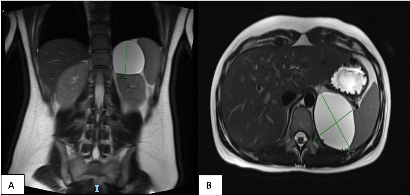
Case Description/Methods: A 29-year-old woman with no significant past medical history presented to her PCP for two weeks of sharp left upper quadrant (LUQ) abdominal pain after a Pure Barre class. It was exacerbated by exertion and improved with rest. Physical exam was notable for tenderness to palpation above the left clavicle and left upper quadrant (LUQ) with no palpable abnormality, CVA tenderness, or splenomegaly. Labs were notable for WBC count 5.1, D-dimer < 0.19, and negative urine analysis. Her presentation was thought to be consistent with musculoskeletal pain and she was advised conservative management. Her pain didn’t improve, and she was ordered for an abdominal ultrasound which showed a LUQ cyst measuring 7.1cm and abutting the upper pole of the left kidney, spleen, and pancreatic tail. MRI abdomen with contrast showed a 7.7 x 6.0 x 6.5cm simple thin-walled cyst with no solid or nodular enhancement, likely reflecting a benign RP cyst. She was referred to urology who recommended a stability scan at 6 months and drainage if her symptoms persisted. Her cyst remained stable in size at 6 months, and she became asymptomatic at the next visit so no invasive interventions were pursued.
Discussion: RP cysts pose an important diagnostic and therapeutic challenge to physicians. They have an estimated incidence of 1/250,000 and cause vague symptoms such as abdominal discomfort and slowly increasing abdominal girth. They are commonly classified as either neoplastic or non-neoplastic and diagnosis is by radiologic imaging or pathology following drainage. The preferred treatment for symptomatic RP cysts is often excision when complications like perforation, infection, or malignancy are expected. However, surgery within the RP space close to major vessels carries an inherent risk of serious complications, and partial excision/drainage is often associated with recurrence. Therefore, a watchful waiting approach for asymptomatic RP cysts with benign characteristics including thin, regular walls, lack of septae, or solid components may be considered as in the case above.

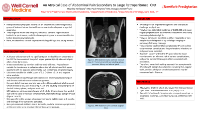
Figure: MRI Abdomen with and without contrast: Coronal (A) and Sagittal (B) Views of 7.7 x 6.0 x 6.5cm RP cyst abutting medial aspect of the spleen, posterior aspect of the pancreas, superior aspect of kidney, and posterior aspect of the adrenal gland as well as diaphragm.
Disclosures:
Haarika Korlipara indicated no relevant financial relationships.
Douglas Scherr indicated no relevant financial relationships.
Paul Fenyves indicated no relevant financial relationships.
Haarika Korlipara, MD1, Douglas Scherr, MD2, Paul Fenyves, MD2. P3770 - An Atypical Case of Abdominal Pain Secondary to Large Retroperitoneal Cyst, ACG 2024 Annual Scientific Meeting Abstracts. Philadelphia, PA: American College of Gastroenterology.
