Tuesday Poster Session
Category: Colon
P3773 - Xanthogranulomatous Appendicitis Mimicking an Appendiceal Malignancy
Tuesday, October 29, 2024
10:30 AM - 4:00 PM ET
Location: Exhibit Hall E

Has Audio
- SK
Sharanya Kumar, MD
Riverside University Health System
Moreno Valley, CA
Presenting Author(s)
Sharanya Kumar, MD1, Jun Song, MD2, Harold Duarte, MD2, Ronaldo Gnass, MD1, Christopher Bent, MD1, Rahul Tuli, BS3, Allen Seylani, BS3, Mena Saad, DO1, Maher Tama, MD1, Steve Serrao, MD1
1Riverside University Health System, Moreno Valley, CA; 2Loma Linda University Medical Center, Moreno Valley, CA; 3University of California Riverside School of Medicine, Riverside, CA
Introduction: Xanthogranulomatous inflammation (XGI) is a rare chronic inflammatory condition that typically affects the kidney and gallbladder, with appendicitis being an exceptionally uncommon manifestation. Radiologically, XGI appears as a destructive mass lesion that mimics malignancy and complicates diagnosis, as described in the case below.
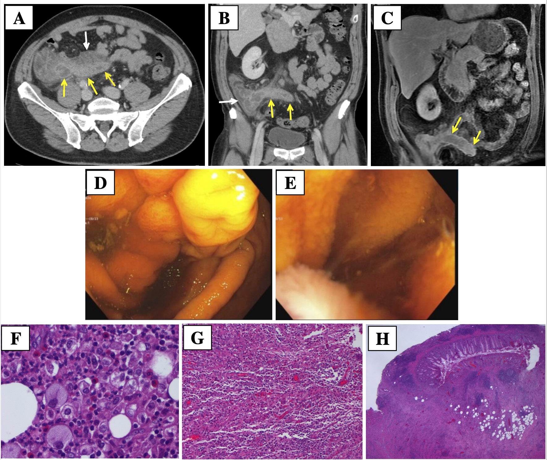
Case Description/Methods: A 55-year-old male with hypertension and ulcerative colitis presented with abdominal pain. Labs revealed leukocytosis, while CEA, CA 19-9, and CA-125 were negative. Abdominal CT showed wall thickening and inflammation of the appendix and cecum with an adjacent soft tissue density and prominent regional lymph nodes (Fig A-B). MRI of the abdomen and pelvis with and without contrast showed scattered regional lymphadenopathy, wall thickening of the distal cecum, and distension of the appendix with regional edema, suggestive of mucinous obstruction (Fig C). Colonoscopy revealed an inflamed and protuberant appendiceal orifice with minimal purulent drainage (Fig D-E). Subsequently, colorectal surgery performed a robotic-assisted right hemicolectomy with primary isoperistaltic ileocolonic anastomosis. Intraoperatively, a dilated and thickened appendix with severe desmoplastic reaction to the sigmoid colon, omentum, mesentery, and retroperitoneum was noted. Histopathology of the right hemicolon and appendix showed xanthogranulomatous appendicitis/periappendicitis with aggregates of foamy histiocytes and macrophages in an inflammatory infiltrate of lymphocytes, plasma cells, and neutrophils, diagnostic for XGI (Fig F-H). After discharge the patient followed up with Surgical Oncology and reported resolution of his symptoms.
Discussion: XGI is a rare histopathologic condition resulting from defective lipid transport, immunologic disturbances, and chronic infections. Diagnosis of XGI is challenging due to its radiologic similarities to malignancy, and it is often identified with histopathology after surgical intervention. This case highlights the importance of obtaining tissue biopsy during diagnostic procedures as it may avoid emergent surgery. Rapid onsite touch imprint cytology may be useful in these cases to help differentiate between benign and malignant cells intraoperatively. Furthermore, endoscopic retrograde appendicitis therapy, a tool involving plastic stent placement, may serve as a bridge to surgery. Overall, further clinical experience with XGI appendicitis is needed to determine the optimal endoscopic treatment modality.

Disclosures:
Sharanya Kumar, MD1, Jun Song, MD2, Harold Duarte, MD2, Ronaldo Gnass, MD1, Christopher Bent, MD1, Rahul Tuli, BS3, Allen Seylani, BS3, Mena Saad, DO1, Maher Tama, MD1, Steve Serrao, MD1. P3773 - Xanthogranulomatous Appendicitis Mimicking an Appendiceal Malignancy, ACG 2024 Annual Scientific Meeting Abstracts. Philadelphia, PA: American College of Gastroenterology.
1Riverside University Health System, Moreno Valley, CA; 2Loma Linda University Medical Center, Moreno Valley, CA; 3University of California Riverside School of Medicine, Riverside, CA
Introduction: Xanthogranulomatous inflammation (XGI) is a rare chronic inflammatory condition that typically affects the kidney and gallbladder, with appendicitis being an exceptionally uncommon manifestation. Radiologically, XGI appears as a destructive mass lesion that mimics malignancy and complicates diagnosis, as described in the case below.
Case Description/Methods: A 55-year-old male with hypertension and ulcerative colitis presented with abdominal pain. Labs revealed leukocytosis, while CEA, CA 19-9, and CA-125 were negative. Abdominal CT showed wall thickening and inflammation of the appendix and cecum with an adjacent soft tissue density and prominent regional lymph nodes (Fig A-B). MRI of the abdomen and pelvis with and without contrast showed scattered regional lymphadenopathy, wall thickening of the distal cecum, and distension of the appendix with regional edema, suggestive of mucinous obstruction (Fig C). Colonoscopy revealed an inflamed and protuberant appendiceal orifice with minimal purulent drainage (Fig D-E). Subsequently, colorectal surgery performed a robotic-assisted right hemicolectomy with primary isoperistaltic ileocolonic anastomosis. Intraoperatively, a dilated and thickened appendix with severe desmoplastic reaction to the sigmoid colon, omentum, mesentery, and retroperitoneum was noted. Histopathology of the right hemicolon and appendix showed xanthogranulomatous appendicitis/periappendicitis with aggregates of foamy histiocytes and macrophages in an inflammatory infiltrate of lymphocytes, plasma cells, and neutrophils, diagnostic for XGI (Fig F-H). After discharge the patient followed up with Surgical Oncology and reported resolution of his symptoms.
Discussion: XGI is a rare histopathologic condition resulting from defective lipid transport, immunologic disturbances, and chronic infections. Diagnosis of XGI is challenging due to its radiologic similarities to malignancy, and it is often identified with histopathology after surgical intervention. This case highlights the importance of obtaining tissue biopsy during diagnostic procedures as it may avoid emergent surgery. Rapid onsite touch imprint cytology may be useful in these cases to help differentiate between benign and malignant cells intraoperatively. Furthermore, endoscopic retrograde appendicitis therapy, a tool involving plastic stent placement, may serve as a bridge to surgery. Overall, further clinical experience with XGI appendicitis is needed to determine the optimal endoscopic treatment modality.

Figure: Figures A & B: CT scan of the abdomen and pelvis with IV contrast in axial view followed by coronal view. Yellow arrows highlight the enlarged appendix, and white arrows indicate inflammatory fluid adjacent to the thickened cecal wall
Figure C: MRI of the abdomen and pelvis with IV contrast in the coronal view, with arrows indicating an enhancing appendix
Figure D-E: Images of the cecum and terminal ileum taken during diagnostic colonoscopy
Figure F-H: Mixed inflammatory infiltrate in an appendiceal biopsy showing foamy histiocytes, plasma cells, neutrophils, and eosinophils, indicative of xanthogranulomatous inflammation (XGI) (400x, 100x, 12.5x)
Disclosures:
Sharanya Kumar indicated no relevant financial relationships.
Jun Song indicated no relevant financial relationships.
Harold Duarte indicated no relevant financial relationships.
Ronaldo Gnass indicated no relevant financial relationships.
Christopher Bent indicated no relevant financial relationships.
Rahul Tuli indicated no relevant financial relationships.
Allen Seylani indicated no relevant financial relationships.
Mena Saad indicated no relevant financial relationships.
Maher Tama indicated no relevant financial relationships.
Steve Serrao: Provation Medical – Advisory Committee/Board Member.
Sharanya Kumar, MD1, Jun Song, MD2, Harold Duarte, MD2, Ronaldo Gnass, MD1, Christopher Bent, MD1, Rahul Tuli, BS3, Allen Seylani, BS3, Mena Saad, DO1, Maher Tama, MD1, Steve Serrao, MD1. P3773 - Xanthogranulomatous Appendicitis Mimicking an Appendiceal Malignancy, ACG 2024 Annual Scientific Meeting Abstracts. Philadelphia, PA: American College of Gastroenterology.

