Tuesday Poster Session
Category: Colon
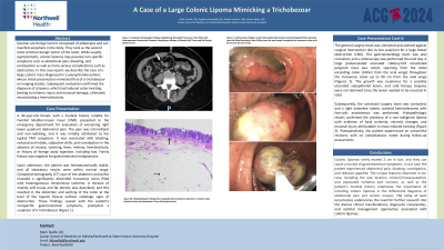
P3775 - A Case of a Large Colonic Lipoma Mimicking a Trichobezoar
Tuesday, October 29, 2024
10:30 AM - 4:00 PM ET
Location: Exhibit Hall E

Has Audio
.jpg)
Mark Tawfik, DO
Staten Island University Hospital, Northwell Health
Staten Island, NY
Presenting Author(s)
Mark Tawfik, DO1, Angelica Rozenfeld, BA2, Malek Kreidieh, MD1, Liliane Deeb, MD1
1Staten Island University Hospital, Northwell Health, Staten Island, NY; 2Lake Erie College of Osteopathic Medicine, Staten Island, NY
Introduction: Colonic lipomas are benign mesenchymal tumors composed of mature adipocytes. They are the second most common benign neoplasms of the colon after adenomatous polyps. They favor the ascending colon, followed by the sigmoid colon, descending colon, and transverse colon. Although colonic lipomas are typically asymptomatic, larger ones can elicit a spectrum of gastrointestinal symptoms, ranging from vague discomfort to more alarming manifestations such as bleeding or obstruction. Interestingly, their propensity for nonspecific presentations often poses a considerable challenge in their diagnosis.
Case Description/Methods: A 36-year-old female with a medical history notable for Familial Mediterranean Fever (FMF) presented to the emergency department
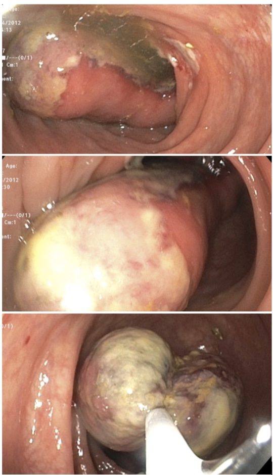
for evaluation of worsening right lower quadrant abdominal pain. Computed tomography (CT) scan of the abdomen and pelvis revealed a significantly distended transverse colon filled with heterogeneous intraluminal contents. A mixture of strandy soft tissue and fat density was described, and this resulted in the distortion and swirling of the colon at the level of the hepatic flexure without radiologic signs of obstruction. These findings prompted a suspicion of a trichobezoar. Gastroenterology was consulted and a colonoscopy was performed the next day. A large pedunculated ulcerated rubbery-felt convoluted polypoid mass was noted, spanning from the distal ascending colon (140cm from the anal verge) throughout the transverse colon up to 90 cm from the anal verge (Figures 1). Subsequently, colorectal surgery team was contacted, and right extended robotic assisted hemicolectomy with ileo-colic anastomosis was performed. Histopathologic results confirmed the presence of a non-malignant lipoma with evidence of focal ischemia, necrotic changes, and mucosal injury attributable to mass-induced twisting. Postoperatively, the patient experienced an uneventful recovery with no complications noted during follow-up assessments.
Discussion: Colonic lipomas rarely exceed 2 cm in size, and they can cause a myriad of gastrointestinal symptoms. In our case the patient experienced abdominal pain, bloating, constipation, and reduced appetite. The unique features observed in our case, including the size, location, torsion/intussusception, and associated ischemia and necrosis, as well as the patient’s medical history emphasize the importance of including colonic lipomas in the differential diagnosis of abdominal pain and colonic masses.

Disclosures:
Mark Tawfik, DO1, Angelica Rozenfeld, BA2, Malek Kreidieh, MD1, Liliane Deeb, MD1. P3775 - A Case of a Large Colonic Lipoma Mimicking a Trichobezoar, ACG 2024 Annual Scientific Meeting Abstracts. Philadelphia, PA: American College of Gastroenterology.
1Staten Island University Hospital, Northwell Health, Staten Island, NY; 2Lake Erie College of Osteopathic Medicine, Staten Island, NY
Introduction: Colonic lipomas are benign mesenchymal tumors composed of mature adipocytes. They are the second most common benign neoplasms of the colon after adenomatous polyps. They favor the ascending colon, followed by the sigmoid colon, descending colon, and transverse colon. Although colonic lipomas are typically asymptomatic, larger ones can elicit a spectrum of gastrointestinal symptoms, ranging from vague discomfort to more alarming manifestations such as bleeding or obstruction. Interestingly, their propensity for nonspecific presentations often poses a considerable challenge in their diagnosis.
Case Description/Methods: A 36-year-old female with a medical history notable for Familial Mediterranean Fever (FMF) presented to the emergency department
for evaluation of worsening right lower quadrant abdominal pain. Computed tomography (CT) scan of the abdomen and pelvis revealed a significantly distended transverse colon filled with heterogeneous intraluminal contents. A mixture of strandy soft tissue and fat density was described, and this resulted in the distortion and swirling of the colon at the level of the hepatic flexure without radiologic signs of obstruction. These findings prompted a suspicion of a trichobezoar. Gastroenterology was consulted and a colonoscopy was performed the next day. A large pedunculated ulcerated rubbery-felt convoluted polypoid mass was noted, spanning from the distal ascending colon (140cm from the anal verge) throughout the transverse colon up to 90 cm from the anal verge (Figures 1). Subsequently, colorectal surgery team was contacted, and right extended robotic assisted hemicolectomy with ileo-colic anastomosis was performed. Histopathologic results confirmed the presence of a non-malignant lipoma with evidence of focal ischemia, necrotic changes, and mucosal injury attributable to mass-induced twisting. Postoperatively, the patient experienced an uneventful recovery with no complications noted during follow-up assessments.
Discussion: Colonic lipomas rarely exceed 2 cm in size, and they can cause a myriad of gastrointestinal symptoms. In our case the patient experienced abdominal pain, bloating, constipation, and reduced appetite. The unique features observed in our case, including the size, location, torsion/intussusception, and associated ischemia and necrosis, as well as the patient’s medical history emphasize the importance of including colonic lipomas in the differential diagnosis of abdominal pain and colonic masses.

Figure: Figure 1: Colonoscopy Findings: Large Pedunculated Ulcerated Convoluted Polypoid Mass Spanning from the Distal Ascending Colon (140cm from the anal verge) throughout the transverse colon up to 90 cm from the anal verge.
Disclosures:
Mark Tawfik indicated no relevant financial relationships.
Angelica Rozenfeld indicated no relevant financial relationships.
Malek Kreidieh indicated no relevant financial relationships.
Liliane Deeb indicated no relevant financial relationships.
Mark Tawfik, DO1, Angelica Rozenfeld, BA2, Malek Kreidieh, MD1, Liliane Deeb, MD1. P3775 - A Case of a Large Colonic Lipoma Mimicking a Trichobezoar, ACG 2024 Annual Scientific Meeting Abstracts. Philadelphia, PA: American College of Gastroenterology.
