Tuesday Poster Session
Category: Colon
P3707 - Vanishing Colonic Mass
Tuesday, October 29, 2024
10:30 AM - 4:00 PM ET
Location: Exhibit Hall E

Has Audio
- AV
Adam Vinall, MD
University of Texas Health Science Center
San Antonio, TX
Presenting Author(s)
Adam Vinall, MD1, Peter Stawinski, MD1, Randy Wright, MD2
1University of Texas Health Science Center, San Antonio, TX; 2University of Texas Health San Antonio, San Antonio, TX
Introduction: Colonoscopy in ischemic colitis most frequently shows mucosal pallor, erythema, and ulceration. Rarely, the ulcerations can be so severe they mimic a colonic mass. This atypical presentation can mimic more common conditions such as colorectal cancer, leading to potential delays in appropriate management. In this case report, we describe a patient with ischemic colitis presenting as a vanishing colonic mass.
Case Description/Methods: A 58-year-old male with hypertension, chronic kidney disease, atrial fibrillation on apixaban, hyperlipidemia, type 2 diabetes mellitus, heart failure with reduced ejection fraction, and alcohol associated cirrhosis, decompensated by ascites, presented to the emergency department after being found down at home with a large amount of hematochezia. On exam, the patient was hemodynamically stable and afebrile. Physical exam was notable for bright red blood on rectal exam, atrial fibrillation, and residual deficits from prior stroke. Labs showed leukocytosis with white blood cell count of 10.9K, hemoglobin of 8.9 g/dL, down from 14.9 g/dL weeks prior, and platelet count of 116K. Renal and liver function tests were stable from prior. Initial lactic acid was 4.3mmol/L.
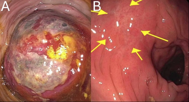
CT angiogram of the abdomen demonstrated a distended rectosigmoid colon filled with blood clots without evidence of active extravasation and hemoperitoneum. Diagnostic colonoscopy showed a large exophytic, subepithelial mass in the rectum, with possible penetration in the peritoneum, as seen in Figure A. Biopsy showed abundant fibrinopurulent ulcer debris and fragments of colonic mucosa with ischemic change. General surgery was consulted and decided against surgical intervention due to the inflammatory appearance of the mass. Follow up colonoscopy eight months later showed complete resolution of the mass with remnant scar, as seen in Figure B.
Discussion: The presentation of ischemic colitis as a colonic mass is a rare and diagnostically challenging phenomenon. Ischemic colitis affects the rectum in only 1% of cases, which made diagnosis more difficult in this patient. Pathology is crucial for obtaining the correct diagnosis, which should demonstrate nonspecific inflammatory changes and absence of neoplasia. Clinicians should consider ischemic colitis in the differential diagnosis of colonic masses, particularly in patients with risk factors such as cardiovascular disease, diabetes, or hypercoagulable states.

Disclosures:
Adam Vinall, MD1, Peter Stawinski, MD1, Randy Wright, MD2. P3707 - Vanishing Colonic Mass, ACG 2024 Annual Scientific Meeting Abstracts. Philadelphia, PA: American College of Gastroenterology.
1University of Texas Health Science Center, San Antonio, TX; 2University of Texas Health San Antonio, San Antonio, TX
Introduction: Colonoscopy in ischemic colitis most frequently shows mucosal pallor, erythema, and ulceration. Rarely, the ulcerations can be so severe they mimic a colonic mass. This atypical presentation can mimic more common conditions such as colorectal cancer, leading to potential delays in appropriate management. In this case report, we describe a patient with ischemic colitis presenting as a vanishing colonic mass.
Case Description/Methods: A 58-year-old male with hypertension, chronic kidney disease, atrial fibrillation on apixaban, hyperlipidemia, type 2 diabetes mellitus, heart failure with reduced ejection fraction, and alcohol associated cirrhosis, decompensated by ascites, presented to the emergency department after being found down at home with a large amount of hematochezia. On exam, the patient was hemodynamically stable and afebrile. Physical exam was notable for bright red blood on rectal exam, atrial fibrillation, and residual deficits from prior stroke. Labs showed leukocytosis with white blood cell count of 10.9K, hemoglobin of 8.9 g/dL, down from 14.9 g/dL weeks prior, and platelet count of 116K. Renal and liver function tests were stable from prior. Initial lactic acid was 4.3mmol/L.
CT angiogram of the abdomen demonstrated a distended rectosigmoid colon filled with blood clots without evidence of active extravasation and hemoperitoneum. Diagnostic colonoscopy showed a large exophytic, subepithelial mass in the rectum, with possible penetration in the peritoneum, as seen in Figure A. Biopsy showed abundant fibrinopurulent ulcer debris and fragments of colonic mucosa with ischemic change. General surgery was consulted and decided against surgical intervention due to the inflammatory appearance of the mass. Follow up colonoscopy eight months later showed complete resolution of the mass with remnant scar, as seen in Figure B.
Discussion: The presentation of ischemic colitis as a colonic mass is a rare and diagnostically challenging phenomenon. Ischemic colitis affects the rectum in only 1% of cases, which made diagnosis more difficult in this patient. Pathology is crucial for obtaining the correct diagnosis, which should demonstrate nonspecific inflammatory changes and absence of neoplasia. Clinicians should consider ischemic colitis in the differential diagnosis of colonic masses, particularly in patients with risk factors such as cardiovascular disease, diabetes, or hypercoagulable states.

Figure: Figure A. Large, necrotic, ischemic ulcer at rectosigmoid junction with mass-like appearance. Biopsy showed fibrinopurulent debris with ischemic change.
Figure B. Colonoscopy eight months later showed complete resolution of the mass, with mucosal scar at the area of the prior mass.
Figure B. Colonoscopy eight months later showed complete resolution of the mass, with mucosal scar at the area of the prior mass.
Disclosures:
Adam Vinall indicated no relevant financial relationships.
Peter Stawinski indicated no relevant financial relationships.
Randy Wright indicated no relevant financial relationships.
Adam Vinall, MD1, Peter Stawinski, MD1, Randy Wright, MD2. P3707 - Vanishing Colonic Mass, ACG 2024 Annual Scientific Meeting Abstracts. Philadelphia, PA: American College of Gastroenterology.
