Tuesday Poster Session
Category: Colon
P3712 - Swirling Strangulated Cecal Volvulus Masked as Small Bowel Obstruction
Tuesday, October 29, 2024
10:30 AM - 4:00 PM ET
Location: Exhibit Hall E

Has Audio

Riddhi Machchhar, DO
Ocean University Medical Center
Brick, NJ
Presenting Author(s)
Riddhi Machchhar, DO, Tejaswi Makkapati, MD, Ambica Nair, MBBS, Stephen Klepner, DO
Ocean University Medical Center, Brick, NJ
Introduction: A 63 year old man had a cecal volvulus misdiagnosed as a small bowel obstruction. He underwent emergency surgery and a right hemicolectomy. Identifying cecal volvulus in cases of suspected small bowel obstruction is crucial to prevent delays in treatment and reduce mortality.
Case Description/Methods: A 63-year-old male presented to the ER with worsening abdominal pain over two days. The patient had a complex medical history, including GI bleed, polyps, internal hemorrhoids, and an exploratory laparotomy following a self-inflicted stab wound to the liver. The exam revealed a severely distended abdomen and absent bowel sounds. Lab results showed leukocytosis, anemia, and elevated creatinine. An abdominal X-ray revealed dilated bowel loops, indicating small bowel obstruction. General Surgery suspected a cecal volvulus due to an observed "whirl" sign on the CT scan and confirmed this during exploratory laparotomy. They performed a right hemicolectomy and removed portions of the terminal ileum. Pathology confirmed mucosal ischemia and necrotic features in the cecum. The patient experienced post-op complications, requiring TPN. He was eventually discharged to rehab.
Discussion: Cecal volvulus is a rare condition in which the intestines become twisted. It was first identified by Rokitansky in 1837. In 2021, it accounted for 1-1.5% of all cases of intestinal obstruction, with an estimated incidence of 2.8-7.1 million cases. It is more common in females at an average age of 61.8 years. Causes include congenital abnormalities or mechanical processes like abdominal procedures, which lead to adhesions, constipation, and immobility. The congenital abnormalities may include incomplete intestinal rotation during embryonic development and laxity of the parietocolic ligament. Symptoms can be mild or acute, including recurrent intermittent symptoms, acute obstruction, and acute fulminant patterns. Diagnosis is critical, as the condition is associated with a high mortality rate. Lab work may reveal leukocytosis and increased C-reactive protein, and imaging studies such as abdominal X-rays and CT scans aid in diagnosis and management. Surgical intervention is the most effective treatment option, including right hemicolectomy, cecum resection with ileocolic anastomosis, manual reduction or detorsion by caeliotomy, and cecopexy. With appropriate surgical intervention before strangulation occurs, the mortality rate of cecal volvulus can be as low as 10-12%, but it increases to 30-40% if strangulation occurs.

Disclosures:
Riddhi Machchhar, DO, Tejaswi Makkapati, MD, Ambica Nair, MBBS, Stephen Klepner, DO. P3712 - Swirling Strangulated Cecal Volvulus Masked as Small Bowel Obstruction, ACG 2024 Annual Scientific Meeting Abstracts. Philadelphia, PA: American College of Gastroenterology.
Ocean University Medical Center, Brick, NJ
Introduction: A 63 year old man had a cecal volvulus misdiagnosed as a small bowel obstruction. He underwent emergency surgery and a right hemicolectomy. Identifying cecal volvulus in cases of suspected small bowel obstruction is crucial to prevent delays in treatment and reduce mortality.
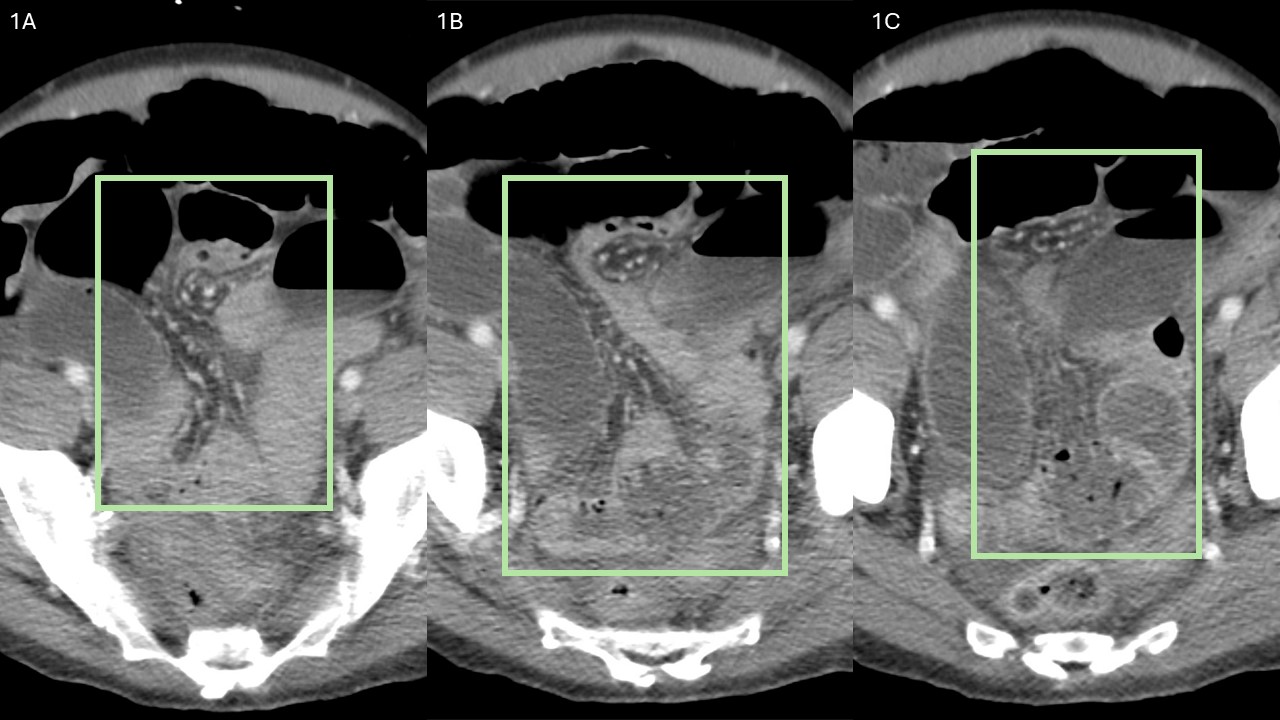
Case Description/Methods: A 63-year-old male presented to the ER with worsening abdominal pain over two days. The patient had a complex medical history, including GI bleed, polyps, internal hemorrhoids, and an exploratory laparotomy following a self-inflicted stab wound to the liver. The exam revealed a severely distended abdomen and absent bowel sounds. Lab results showed leukocytosis, anemia, and elevated creatinine. An abdominal X-ray revealed dilated bowel loops, indicating small bowel obstruction. General Surgery suspected a cecal volvulus due to an observed "whirl" sign on the CT scan and confirmed this during exploratory laparotomy. They performed a right hemicolectomy and removed portions of the terminal ileum. Pathology confirmed mucosal ischemia and necrotic features in the cecum. The patient experienced post-op complications, requiring TPN. He was eventually discharged to rehab.
Discussion: Cecal volvulus is a rare condition in which the intestines become twisted. It was first identified by Rokitansky in 1837. In 2021, it accounted for 1-1.5% of all cases of intestinal obstruction, with an estimated incidence of 2.8-7.1 million cases. It is more common in females at an average age of 61.8 years. Causes include congenital abnormalities or mechanical processes like abdominal procedures, which lead to adhesions, constipation, and immobility. The congenital abnormalities may include incomplete intestinal rotation during embryonic development and laxity of the parietocolic ligament. Symptoms can be mild or acute, including recurrent intermittent symptoms, acute obstruction, and acute fulminant patterns. Diagnosis is critical, as the condition is associated with a high mortality rate. Lab work may reveal leukocytosis and increased C-reactive protein, and imaging studies such as abdominal X-rays and CT scans aid in diagnosis and management. Surgical intervention is the most effective treatment option, including right hemicolectomy, cecum resection with ileocolic anastomosis, manual reduction or detorsion by caeliotomy, and cecopexy. With appropriate surgical intervention before strangulation occurs, the mortality rate of cecal volvulus can be as low as 10-12%, but it increases to 30-40% if strangulation occurs.

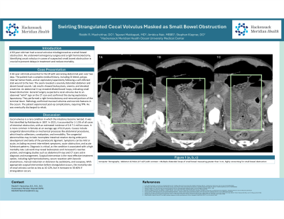
Figure: FIGURE 1 (a, b, c): CT A/P w/ contrast = Multiple distended loops of small bowel measuring greater than 3 cm, highly concerning for small bowel obstruction
Disclosures:
Riddhi Machchhar indicated no relevant financial relationships.
Tejaswi Makkapati indicated no relevant financial relationships.
Ambica Nair indicated no relevant financial relationships.
Stephen Klepner indicated no relevant financial relationships.
Riddhi Machchhar, DO, Tejaswi Makkapati, MD, Ambica Nair, MBBS, Stephen Klepner, DO. P3712 - Swirling Strangulated Cecal Volvulus Masked as Small Bowel Obstruction, ACG 2024 Annual Scientific Meeting Abstracts. Philadelphia, PA: American College of Gastroenterology.
