Tuesday Poster Session
Category: Biliary/Pancreas
P3589 - Diagnostic Challenges in Differentiating Pancreatic Paragangliomas From Well Differentiated Neuroendocrine Tumors
Tuesday, October 29, 2024
10:30 AM - 4:00 PM ET
Location: Exhibit Hall E

Has Audio

Ahmed Fares, MD
Tufts Medical Center
Boston, MA
Presenting Author(s)
Ahmed Fares, MD, Erik Holzwanger, MD, Michael Talanian, MD
Tufts Medical Center, Boston, MA
Introduction: Paragangliomas (PGLs) are rare neuroendocrine tumors arising from extra-adrenal neuroendocrine cells, classified based on their sympathetic or parasympathetic origin. Sympathetic PGLs often present with symptoms similar to pheochromocytomas, whereas parasympathetic PGLs are usually non-secretory and found in the head and neck. Neuroendocrine tumors (NETs) represent a diverse group of neoplasms that can secrete various hormones, leading to a range of clinical manifestations. This case report discusses a 58-year-old female with a pancreatic mass, highlighting the diagnostic challenges in differentiating between PGLs and well-differentiated NETs.
Case Description/Methods: A 58-year-old female with a history of diverticulitis and sigmoidectomy presented with vomiting, diarrhea, and abdominal pain. An ultrasound revealed a hypoechoic lesion in the pancreatic head and a stable adrenal mass first detected 16 years previously. Both were also seen on computed tomography (figure 1). Endoscopic ultrasound (EUS) identified a mass in the body of the pancreas. Biopsy was positive for neoplastic cells favoring PGL, with immunohistochemical staining positive for chromogranin and synaptophysin, and negative for pankeratin, CAM5.2, S100, GATA3, tyrosine hydroxylase, and beta-catenin. Catecholamine and neuroendocrine marker tests were normal (table 1). The absence of typical PGL symptoms and negative catecholamines suggested a non-secretory PGL or NET.
Discussion: Differentiating PGLs from NETs is complex due to overlapping clinical and immunohistochemical features. In this case, immunohistochemical staining was critical, though presence of markers common to both PGLs and NETs made the diagnosis difficult. Molecular analysis, including microRNA expression profiles, has been outlined by studies to provide additional diagnostic criteria, although not performed in this case given limited sampling. The case underscores the importance of thorough diagnostic workups and pathological assessments to differentiate between the two groups of tumors and guide appropriate treatment. Surgical resection remains the treatment of choice for localized PGLs, with a focus on detecting metastases given their potential for malignancy.

Note: The table for this abstract can be viewed in the ePoster Gallery section of the ACG 2024 ePoster Site or in The American Journal of Gastroenterology's abstract supplement issue, both of which will be available starting October 27, 2024.
Disclosures:
Ahmed Fares, MD, Erik Holzwanger, MD, Michael Talanian, MD. P3589 - Diagnostic Challenges in Differentiating Pancreatic Paragangliomas From Well Differentiated Neuroendocrine Tumors, ACG 2024 Annual Scientific Meeting Abstracts. Philadelphia, PA: American College of Gastroenterology.
Tufts Medical Center, Boston, MA
Introduction: Paragangliomas (PGLs) are rare neuroendocrine tumors arising from extra-adrenal neuroendocrine cells, classified based on their sympathetic or parasympathetic origin. Sympathetic PGLs often present with symptoms similar to pheochromocytomas, whereas parasympathetic PGLs are usually non-secretory and found in the head and neck. Neuroendocrine tumors (NETs) represent a diverse group of neoplasms that can secrete various hormones, leading to a range of clinical manifestations. This case report discusses a 58-year-old female with a pancreatic mass, highlighting the diagnostic challenges in differentiating between PGLs and well-differentiated NETs.
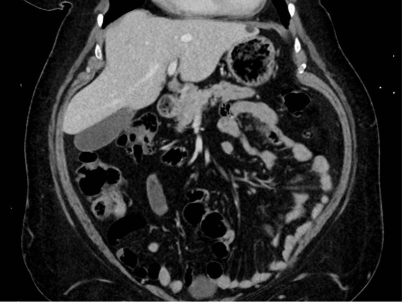
Case Description/Methods: A 58-year-old female with a history of diverticulitis and sigmoidectomy presented with vomiting, diarrhea, and abdominal pain. An ultrasound revealed a hypoechoic lesion in the pancreatic head and a stable adrenal mass first detected 16 years previously. Both were also seen on computed tomography (figure 1). Endoscopic ultrasound (EUS) identified a mass in the body of the pancreas. Biopsy was positive for neoplastic cells favoring PGL, with immunohistochemical staining positive for chromogranin and synaptophysin, and negative for pankeratin, CAM5.2, S100, GATA3, tyrosine hydroxylase, and beta-catenin. Catecholamine and neuroendocrine marker tests were normal (table 1). The absence of typical PGL symptoms and negative catecholamines suggested a non-secretory PGL or NET.
Discussion: Differentiating PGLs from NETs is complex due to overlapping clinical and immunohistochemical features. In this case, immunohistochemical staining was critical, though presence of markers common to both PGLs and NETs made the diagnosis difficult. Molecular analysis, including microRNA expression profiles, has been outlined by studies to provide additional diagnostic criteria, although not performed in this case given limited sampling. The case underscores the importance of thorough diagnostic workups and pathological assessments to differentiate between the two groups of tumors and guide appropriate treatment. Surgical resection remains the treatment of choice for localized PGLs, with a focus on detecting metastases given their potential for malignancy.

Figure: Figure 1: CT scan demonstrated a 1.2 cm enhancing lesion in the body of the pancreas.
Note: The table for this abstract can be viewed in the ePoster Gallery section of the ACG 2024 ePoster Site or in The American Journal of Gastroenterology's abstract supplement issue, both of which will be available starting October 27, 2024.
Disclosures:
Ahmed Fares indicated no relevant financial relationships.
Erik Holzwanger: Boston Scientific – Consultant.
Michael Talanian indicated no relevant financial relationships.
Ahmed Fares, MD, Erik Holzwanger, MD, Michael Talanian, MD. P3589 - Diagnostic Challenges in Differentiating Pancreatic Paragangliomas From Well Differentiated Neuroendocrine Tumors, ACG 2024 Annual Scientific Meeting Abstracts. Philadelphia, PA: American College of Gastroenterology.
