Tuesday Poster Session
Category: Biliary/Pancreas
P3590 - Third Time's the Charm? An Elusive Case of Pancreatic Rosai-Dorfman Disease
Tuesday, October 29, 2024
10:30 AM - 4:00 PM ET
Location: Exhibit Hall E

Has Audio
- GS
Gurdeep Singh, DO
AdventHealth
Orlando, FL
Presenting Author(s)
Gurdeep Singh, DO1, William Cobb, MD1, Haven Ward, MD1, Arooj Mian, MD2, Karen Echeverria-Beltran, MD1, Mustafa Arain, MD1, Armando Rosales, MD1
1AdventHealth, Orlando, FL; 2AdventHealth Medical Group, AdventHealth, Orlando, FL
Introduction: Rosai-Dorfman Disease (RDD) is a benign, proliferative disease of non-Langerhans cell histiocytes typically involving lymphoid tissue that was first described in 1965 and further classified in 1969. RDD is rare and cases of extra-nodal disease involving the skin, lungs, liver, eyes, nervous system, and kidneys have been reported. Pancreatic involvement is extremely rare.
Case Description/Methods: A 53-year-old female who originally presented with vague epigastric abdominal pain. Magnetic resonance imaging (MRI) revealed a hypoechoic lesion in the proximal pancreatic body concerning for a mass. Endoscopic ultrasound (EUS) was consistent with a round, hypoechoic mass in the pancreatic body. Fine needle aspiration (FNA) demonstrated features of acute and chronic inflammation in a background of extensive fibrosis with no malignancy. Repeat EUS-FNA performed two months later was also negative for malignancy. Initially, the patient stated she did not want to undergo surgery and preferred conservative management.
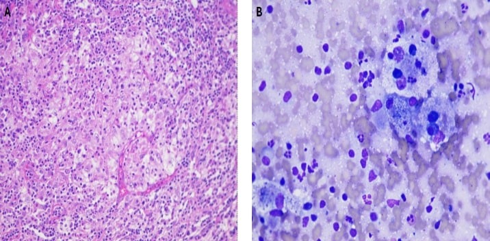
She was monitored closely and underwent a surveillance MRI of the abdomen four months later that showed interval growth of the pancreatic lesion from 2.5 x 2.1 cm to 2.9 x 3.0 cm. After repeated discussion, the patient successfully underwent a robotic-assisted subtotal pancreatectomy and splenectomy. The final pathology revealed sheets of foamy histiocytes with enlarged vesicular nuclei and nucleoli and numerous neutrophils, lymphocytes, and plasma cells. Immunohistochemical testing was positive for S-100 protein and CD163 and negative for CD1a and ALK-1, supporting a diagnosis of Rosai-Dorfman Disease. Next generation sequencing was positive for an F53L variant mutation of the MAP2K1 gene. The patient made an uneventful recovery from surgery and remains asymptomatic with a plan for repeat MRI imaging 6 months later.
Discussion: Diagnosing RDD can often be challenging as it hinges on obtaining an adequate tissue sample for histopathologic analysis. The key cytomorphologic feature is “emperipolesis,” the engulfment of lymphocytes, plasma cells, and occasionally, erythrocytes by histocytes without lysosomal degradation.
Immunophenotyping studies for RDD are typically positive for S100 protein, CD68, CD163, and OCT2 and negative for CD1a and CD207 (Langerhans cell markers). Overall, there is no uniform approach for the treatment of RDD and is usually tailored to the individual’s clinical picture.

Disclosures:
Gurdeep Singh, DO1, William Cobb, MD1, Haven Ward, MD1, Arooj Mian, MD2, Karen Echeverria-Beltran, MD1, Mustafa Arain, MD1, Armando Rosales, MD1. P3590 - Third Time's the Charm? An Elusive Case of Pancreatic Rosai-Dorfman Disease, ACG 2024 Annual Scientific Meeting Abstracts. Philadelphia, PA: American College of Gastroenterology.
1AdventHealth, Orlando, FL; 2AdventHealth Medical Group, AdventHealth, Orlando, FL
Introduction: Rosai-Dorfman Disease (RDD) is a benign, proliferative disease of non-Langerhans cell histiocytes typically involving lymphoid tissue that was first described in 1965 and further classified in 1969. RDD is rare and cases of extra-nodal disease involving the skin, lungs, liver, eyes, nervous system, and kidneys have been reported. Pancreatic involvement is extremely rare.
Case Description/Methods: A 53-year-old female who originally presented with vague epigastric abdominal pain. Magnetic resonance imaging (MRI) revealed a hypoechoic lesion in the proximal pancreatic body concerning for a mass. Endoscopic ultrasound (EUS) was consistent with a round, hypoechoic mass in the pancreatic body. Fine needle aspiration (FNA) demonstrated features of acute and chronic inflammation in a background of extensive fibrosis with no malignancy. Repeat EUS-FNA performed two months later was also negative for malignancy. Initially, the patient stated she did not want to undergo surgery and preferred conservative management.
She was monitored closely and underwent a surveillance MRI of the abdomen four months later that showed interval growth of the pancreatic lesion from 2.5 x 2.1 cm to 2.9 x 3.0 cm. After repeated discussion, the patient successfully underwent a robotic-assisted subtotal pancreatectomy and splenectomy. The final pathology revealed sheets of foamy histiocytes with enlarged vesicular nuclei and nucleoli and numerous neutrophils, lymphocytes, and plasma cells. Immunohistochemical testing was positive for S-100 protein and CD163 and negative for CD1a and ALK-1, supporting a diagnosis of Rosai-Dorfman Disease. Next generation sequencing was positive for an F53L variant mutation of the MAP2K1 gene. The patient made an uneventful recovery from surgery and remains asymptomatic with a plan for repeat MRI imaging 6 months later.
Discussion: Diagnosing RDD can often be challenging as it hinges on obtaining an adequate tissue sample for histopathologic analysis. The key cytomorphologic feature is “emperipolesis,” the engulfment of lymphocytes, plasma cells, and occasionally, erythrocytes by histocytes without lysosomal degradation.
Immunophenotyping studies for RDD are typically positive for S100 protein, CD68, CD163, and OCT2 and negative for CD1a and CD207 (Langerhans cell markers). Overall, there is no uniform approach for the treatment of RDD and is usually tailored to the individual’s clinical picture.

Figure: H&E slides depicting sheets of large foamy neoplastic cells admixed with various inflammatory cells, noting the presence of inflammatory cells within the cytoplasm of neoplastic cells (“emperipolesis”) at 200x magnification (A) and 400x magnification (B).
Disclosures:
Gurdeep Singh indicated no relevant financial relationships.
William Cobb indicated no relevant financial relationships.
Haven Ward indicated no relevant financial relationships.
Arooj Mian indicated no relevant financial relationships.
Karen Echeverria-Beltran indicated no relevant financial relationships.
Mustafa Arain indicated no relevant financial relationships.
Armando Rosales indicated no relevant financial relationships.
Gurdeep Singh, DO1, William Cobb, MD1, Haven Ward, MD1, Arooj Mian, MD2, Karen Echeverria-Beltran, MD1, Mustafa Arain, MD1, Armando Rosales, MD1. P3590 - Third Time's the Charm? An Elusive Case of Pancreatic Rosai-Dorfman Disease, ACG 2024 Annual Scientific Meeting Abstracts. Philadelphia, PA: American College of Gastroenterology.
