Tuesday Poster Session
Category: Biliary/Pancreas
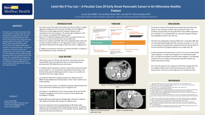
P3505 - Catch Me If You Can: A Peculiar Case of Early Onset Pancreatic Cancer in an Otherwise Healthy Patient
Tuesday, October 29, 2024
10:30 AM - 4:00 PM ET
Location: Exhibit Hall E


Usman Afzal, MD
MedStar Georgetown University Hospital
Washington, DC
Presenting Author(s)
Usman Afzal, MD1, Ahmad Abdulraheem, MD1, Rabi Lee, NP2, Alireza Meighani, MD3, Sayel Alzraikat, MD4
1MedStar Georgetown University Hospital, Washington, DC; 2Medstar Washington Hospital Center, Washington, DC; 3MedStar Health, Washington Hospital Center, Washington, DC; 4MedStar Health-Georgetown/Washington Hospital Center, Washington, DC
Introduction: Pancreatic cancer (Pancreatic Ductal Adenocarcinoma, PDAC) is a highly aggressive malignancy with a five-year mortality rate of 97-98% (1). Nearly 5.7% of the patients diagnosed with Pancreatic cancer are under age 50 years, referred to as early-onset pancreatic cancer (EOPC) (4). Outcomes in patients with EOPC tend to be marginally better, however, prognosis is still heavily influenced by the stage at which the cancer is diagnosed.(5–7) Smoking and genetics are the biggest risk factors associated with it (4).
Case Description/Methods: We present a case of a 28-year-old male with a pancreatic mass that required ERCP with stent placement from an outside hospital who presented with RUQ abdominal pain and nausea. CT abdomen pelvis showed two pancreatic masses in pancreatic head and uncinate process (Figure 1). He consumed an unspecified amount of beer and denied having alcohol-related pancreatitis. He was a non-smoker and denied the use of any drugs of abuse.
The patient underwent ERCP with cytology suspicious for adenocarcinoma. MRCP showed additional enhancing lesion in liver. Tumor markers were mildly elevated. Due to inconclusive results, a CT-guided liver biopsy showed benign liver tissue with chronic inflammation, but no malignant cells. EUS identified the mass in the pancreatic head, but the FNA revealed no malignant cells. He was in and out of the hospital with unchanged CT findings.
Repeat ERCP was performed and cytology again showed no malignant cells. Repeat EUS guided FNA showed no malignant cells. Per tumor board discussions CT guided biopsy of RP lymph nodes confirmed poorly differentiated adenocarcinoma. Given the metastatic nature of disease he was recommended chemotherapy and advised outpatient follow-up with oncology; however, since the patient was uninsured, he was lost to follow-up. Further genetic testing was not performed.
Discussion: Clinically and radiologically suspected PDAC that is resectable (IIIB) does not need pathological diagnosis before resection. However, in our case, since the patient presented with suspected metastatic disease histological diagnosis was attempted (6).
The patient underwent EUS guided FNA with biopsy on two occasions with inconclusive results. The sensitivity and specificity of EUS guided FNA is 90 and 98% respectively (7), therefore, the probability of our case had 2 negative biopsies was less than 1%.
This age at diagnosis and the diagnostic challenges faced during the hospitalization make this case valuable addition to the literature.

Disclosures:
Usman Afzal, MD1, Ahmad Abdulraheem, MD1, Rabi Lee, NP2, Alireza Meighani, MD3, Sayel Alzraikat, MD4. P3505 - Catch Me If You Can: A Peculiar Case of Early Onset Pancreatic Cancer in an Otherwise Healthy Patient, ACG 2024 Annual Scientific Meeting Abstracts. Philadelphia, PA: American College of Gastroenterology.
1MedStar Georgetown University Hospital, Washington, DC; 2Medstar Washington Hospital Center, Washington, DC; 3MedStar Health, Washington Hospital Center, Washington, DC; 4MedStar Health-Georgetown/Washington Hospital Center, Washington, DC
Introduction: Pancreatic cancer (Pancreatic Ductal Adenocarcinoma, PDAC) is a highly aggressive malignancy with a five-year mortality rate of 97-98% (1). Nearly 5.7% of the patients diagnosed with Pancreatic cancer are under age 50 years, referred to as early-onset pancreatic cancer (EOPC) (4). Outcomes in patients with EOPC tend to be marginally better, however, prognosis is still heavily influenced by the stage at which the cancer is diagnosed.(5–7) Smoking and genetics are the biggest risk factors associated with it (4).
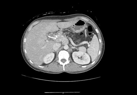
Case Description/Methods: We present a case of a 28-year-old male with a pancreatic mass that required ERCP with stent placement from an outside hospital who presented with RUQ abdominal pain and nausea. CT abdomen pelvis showed two pancreatic masses in pancreatic head and uncinate process (Figure 1). He consumed an unspecified amount of beer and denied having alcohol-related pancreatitis. He was a non-smoker and denied the use of any drugs of abuse.
The patient underwent ERCP with cytology suspicious for adenocarcinoma. MRCP showed additional enhancing lesion in liver. Tumor markers were mildly elevated. Due to inconclusive results, a CT-guided liver biopsy showed benign liver tissue with chronic inflammation, but no malignant cells. EUS identified the mass in the pancreatic head, but the FNA revealed no malignant cells. He was in and out of the hospital with unchanged CT findings.
Repeat ERCP was performed and cytology again showed no malignant cells. Repeat EUS guided FNA showed no malignant cells. Per tumor board discussions CT guided biopsy of RP lymph nodes confirmed poorly differentiated adenocarcinoma. Given the metastatic nature of disease he was recommended chemotherapy and advised outpatient follow-up with oncology; however, since the patient was uninsured, he was lost to follow-up. Further genetic testing was not performed.
Discussion: Clinically and radiologically suspected PDAC that is resectable (IIIB) does not need pathological diagnosis before resection. However, in our case, since the patient presented with suspected metastatic disease histological diagnosis was attempted (6).
The patient underwent EUS guided FNA with biopsy on two occasions with inconclusive results. The sensitivity and specificity of EUS guided FNA is 90 and 98% respectively (7), therefore, the probability of our case had 2 negative biopsies was less than 1%.
This age at diagnosis and the diagnostic challenges faced during the hospitalization make this case valuable addition to the literature.

Figure: CT imaging findings of the pancreatic head mass
Disclosures:
Usman Afzal indicated no relevant financial relationships.
Ahmad Abdulraheem indicated no relevant financial relationships.
Rabi Lee indicated no relevant financial relationships.
Alireza Meighani indicated no relevant financial relationships.
Sayel Alzraikat indicated no relevant financial relationships.
Usman Afzal, MD1, Ahmad Abdulraheem, MD1, Rabi Lee, NP2, Alireza Meighani, MD3, Sayel Alzraikat, MD4. P3505 - Catch Me If You Can: A Peculiar Case of Early Onset Pancreatic Cancer in an Otherwise Healthy Patient, ACG 2024 Annual Scientific Meeting Abstracts. Philadelphia, PA: American College of Gastroenterology.
