Tuesday Poster Session
Category: Biliary/Pancreas
P3506 - Hidden Behind Pancreatitis: Discovering a Solid Pseudopapillary Neoplasm in an Adolescent Male
Tuesday, October 29, 2024
10:30 AM - 4:00 PM ET
Location: Exhibit Hall E

Has Audio

Hiba Khan, MD
Virginia Commonwealth University Health System
Lake Grove, NY
Presenting Author(s)
Hiba Khan, MD1, Mahum Nadeem, MBBS2, Dan McEntire, MD3, Bryce S. Hatfield, MD4, Ravi Vachhani, MD4
1Virginia Commonwealth University Health System, Lake Grove, NY; 2Virginia Commonwealth University Health System, Henrico, VA; 3Virginia Commonwealth University Medical Center, Richmond, VA; 4Virginia Commonwealth University Health System, Richmond, VA
Introduction: Solid pseudopapillary neoplasm (SPN) is a rare pancreatic tumor accounting for 1-3% of all pancreatic tumors. SPNs are slow-growing with low malignant potential and are predominantly diagnosed in females with a female-to-male ratio of 10:1. They most commonly occur in the 2nd or 3rd decade of life. The most common presenting symptom is abdominal pain and is present in up to 45% of patients at diagnosis. The standard treatment is surgical resection to mitigate the risk of malignancy. To date, there have been only 8 published cases of acute pancreatitis leading to a SPN diagnosis. Of these, only two cases have been in men, both greater than 30 years of age. This case presents a rare instance of SPN in an adolescent male initially diagnosed with acute pancreatitis.
Case Description/Methods: A 17-year-old Caucasian male with no significant past medical history presented with a three-month history of constant epigastric abdominal pain with radiation to the back. He denied any history of alcohol use, gallstones, or medication use. Lab work showed a normal blood count, calcium level, liver function tests and a lipase of 1397 units/liter. Based on the characteristic abdominal pain and lipase level three times the upper limit of normal, the patient was diagnosed with acute pancreatitis and started on intravenous fluids. Computed tomography (CT) imaging of the abdomen demonstrated a heterogeneous pancreatic head mass. Magnetic resonance imaging confirmed a 2.6 x 3.5 x 2.9 cm solid mass in the pancreatic head with normal caliber pancreatic duct. Fine needle biopsy performed through an upper endoscopic ultrasound (EUS) showed SPN. The patient then underwent a robotic assisted pancreaticoduodenectomy (whipple procedure). The patient reported resolution of pain following the surgery.
Discussion: This case highlights a rare presentation of SPN in an adolescent male. Male patients with SPN account for less than 10% of cases, and rarely SPN presents in teenage males with acute pancreatitis as the presenting symptom. Potential mechanisms for pancreatitis in SPN include ductal obstruction, compression, localized ischemia, and inflammatory response. This case highlights the importance of thorough diagnostic evaluation in atypical presentations of pancreatitis and the utility of advanced imaging and biopsy techniques. Clinicians should consider SPN in the differential diagnosis of pancreatic masses, even in unusual patient demographics and presentations.

Disclosures:
Hiba Khan, MD1, Mahum Nadeem, MBBS2, Dan McEntire, MD3, Bryce S. Hatfield, MD4, Ravi Vachhani, MD4. P3506 - Hidden Behind Pancreatitis: Discovering a Solid Pseudopapillary Neoplasm in an Adolescent Male, ACG 2024 Annual Scientific Meeting Abstracts. Philadelphia, PA: American College of Gastroenterology.
1Virginia Commonwealth University Health System, Lake Grove, NY; 2Virginia Commonwealth University Health System, Henrico, VA; 3Virginia Commonwealth University Medical Center, Richmond, VA; 4Virginia Commonwealth University Health System, Richmond, VA
Introduction: Solid pseudopapillary neoplasm (SPN) is a rare pancreatic tumor accounting for 1-3% of all pancreatic tumors. SPNs are slow-growing with low malignant potential and are predominantly diagnosed in females with a female-to-male ratio of 10:1. They most commonly occur in the 2nd or 3rd decade of life. The most common presenting symptom is abdominal pain and is present in up to 45% of patients at diagnosis. The standard treatment is surgical resection to mitigate the risk of malignancy. To date, there have been only 8 published cases of acute pancreatitis leading to a SPN diagnosis. Of these, only two cases have been in men, both greater than 30 years of age. This case presents a rare instance of SPN in an adolescent male initially diagnosed with acute pancreatitis.
Case Description/Methods: A 17-year-old Caucasian male with no significant past medical history presented with a three-month history of constant epigastric abdominal pain with radiation to the back. He denied any history of alcohol use, gallstones, or medication use. Lab work showed a normal blood count, calcium level, liver function tests and a lipase of 1397 units/liter. Based on the characteristic abdominal pain and lipase level three times the upper limit of normal, the patient was diagnosed with acute pancreatitis and started on intravenous fluids. Computed tomography (CT) imaging of the abdomen demonstrated a heterogeneous pancreatic head mass. Magnetic resonance imaging confirmed a 2.6 x 3.5 x 2.9 cm solid mass in the pancreatic head with normal caliber pancreatic duct. Fine needle biopsy performed through an upper endoscopic ultrasound (EUS) showed SPN. The patient then underwent a robotic assisted pancreaticoduodenectomy (whipple procedure). The patient reported resolution of pain following the surgery.
Discussion: This case highlights a rare presentation of SPN in an adolescent male. Male patients with SPN account for less than 10% of cases, and rarely SPN presents in teenage males with acute pancreatitis as the presenting symptom. Potential mechanisms for pancreatitis in SPN include ductal obstruction, compression, localized ischemia, and inflammatory response. This case highlights the importance of thorough diagnostic evaluation in atypical presentations of pancreatitis and the utility of advanced imaging and biopsy techniques. Clinicians should consider SPN in the differential diagnosis of pancreatic masses, even in unusual patient demographics and presentations.

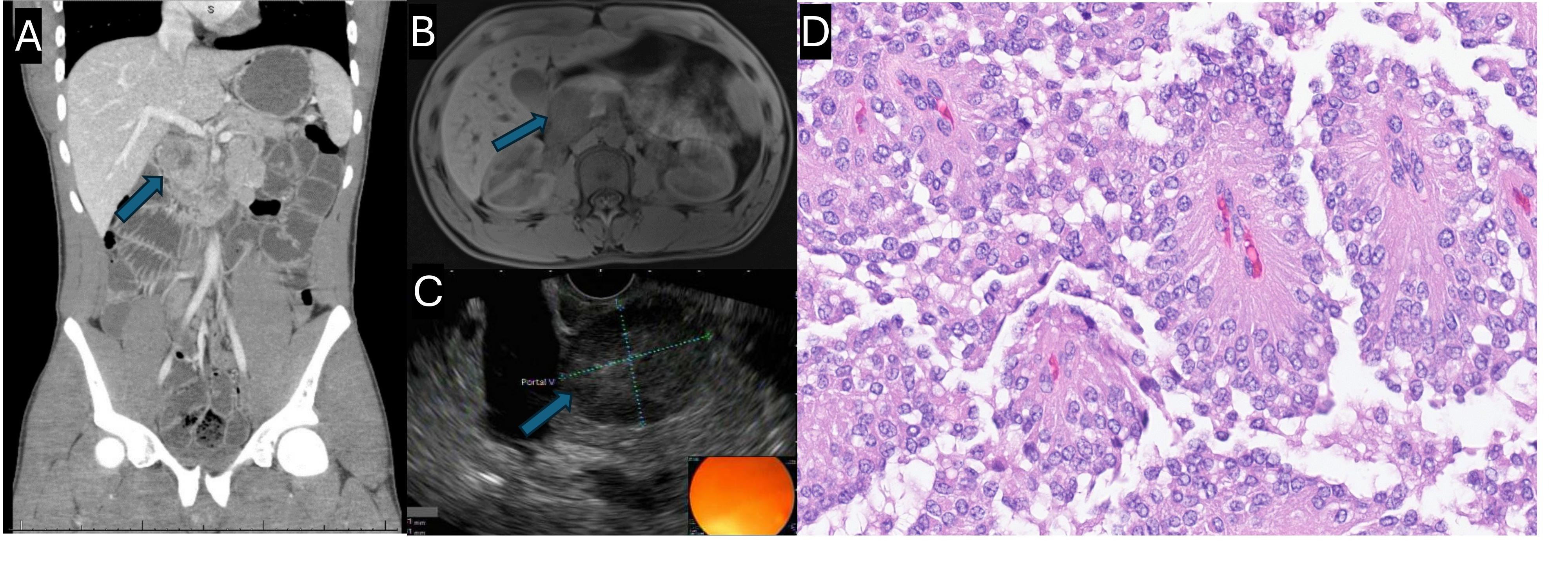
Figure: (A) Computed tomography image of the abdomen and pelvis with arrow pointing to a pancreatic head lesion
(B) Magnetic resonance imaging of the abdomen with arrow pointing to a pancreatic head lesion
(C) Endoscopic ultrasound image showing a pancreatic head lesion
(D) High power microscope image (400x) showing a pseudopapillary appearance with a central fibrovascular-like core, surrounding neoplastic cells with monotonous nuclei, and degenerative spaces between pseudopapillae.
Disclosures:
Hiba Khan indicated no relevant financial relationships.
Mahum Nadeem indicated no relevant financial relationships.
Dan McEntire indicated no relevant financial relationships.
Bryce Hatfield indicated no relevant financial relationships.
Ravi Vachhani indicated no relevant financial relationships.
Hiba Khan, MD1, Mahum Nadeem, MBBS2, Dan McEntire, MD3, Bryce S. Hatfield, MD4, Ravi Vachhani, MD4. P3506 - Hidden Behind Pancreatitis: Discovering a Solid Pseudopapillary Neoplasm in an Adolescent Male, ACG 2024 Annual Scientific Meeting Abstracts. Philadelphia, PA: American College of Gastroenterology.
