Tuesday Poster Session
Category: Biliary/Pancreas
P3511 - Giant Impacted Pancreatic Duct Stone at the Ampulla of Vater
Tuesday, October 29, 2024
10:30 AM - 4:00 PM ET
Location: Exhibit Hall E

Has Audio

Omar Calderon, Jr., MD
University of Wisconsin Hospitals and Clinics
Madison, WI
Presenting Author(s)
Omar Calderon, MD, Eric Vecchio, MD, Anurag Soni, MD, Deepak V. Gopal, MD, FACG
University of Wisconsin Hospitals and Clinics, Madison, WI
Introduction: We present an interesting case of recurrent idiopathic pancreatitis and severe abdominal pain in a patient with a giant pancreatic duct stone obstructing the ampulla of Vater.
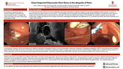
Case Description/Methods: A 74-year-old male with a history of recurrent idiopathic pancreatitis presented to a referring hospital for acute severe epigastric abdominal pain. He denied a history of alcohol or tobacco use for over 20 years. In the emergency room, the patient was hemodynamically stable and afebrile. His abdominal exam was non-tender with normoactive bowel sounds and was non-distended. His labs were significant for lipase 2,108 units/liter. His complete metabolic panel was within normal limits. Imaging the abdomen with magnetic resonance cholangiopancreatography was significant for a 12-millimeter pancreatic head stone contacting and impacted the margin of the ampulla. An endoscopic ultrasound (EUS) and endoscopic retrograde cholangiopancreatography (ERCP) were performed and revealed sequelae of chronic pancreatitis with an impacted large pancreatic duct stone. A pancreatic duct sphincterotomy was performed, followed by balloon sweeps using a triple lumen stone retrieval balloon catheter with successful PD stone removal. A 7 French pancreatic duct stent was placed to prevent post-procedure pancreatitis. The patient’s abdominal pain significantly improved, and he remained symptom free on follow up clinic visit.
Discussion: Patients with chronic pancreatitis and persistent inflammation commonly develop pancreatic calculi with prevalence as high as 60%. These pancreatic duct stones can lead to significant abdominal pain due to obstruction of the pancreatic duct system. Guidelines recommend performing an ERCP with standard stone extraction methods such as sphincterotomy, dilation, and balloon retrieval of the stone. ERCP is typically successful for stones smaller than 5 mm. For stones larger than 5 mm, extracorporeal shock wave lithotripsy (ESWL) is greater than 90% effective at fragmentation, and complete clearance is achieved with ERCP in up to two-thirds of patients following ESWL. In our patient, the stone was impacted at the ampulla and successfully fragmented with sphincterotomy and removed with balloon extraction.

Disclosures:
Omar Calderon, MD, Eric Vecchio, MD, Anurag Soni, MD, Deepak V. Gopal, MD, FACG. P3511 - Giant Impacted Pancreatic Duct Stone at the Ampulla of Vater, ACG 2024 Annual Scientific Meeting Abstracts. Philadelphia, PA: American College of Gastroenterology.
University of Wisconsin Hospitals and Clinics, Madison, WI
Introduction: We present an interesting case of recurrent idiopathic pancreatitis and severe abdominal pain in a patient with a giant pancreatic duct stone obstructing the ampulla of Vater.
Case Description/Methods: A 74-year-old male with a history of recurrent idiopathic pancreatitis presented to a referring hospital for acute severe epigastric abdominal pain. He denied a history of alcohol or tobacco use for over 20 years. In the emergency room, the patient was hemodynamically stable and afebrile. His abdominal exam was non-tender with normoactive bowel sounds and was non-distended. His labs were significant for lipase 2,108 units/liter. His complete metabolic panel was within normal limits. Imaging the abdomen with magnetic resonance cholangiopancreatography was significant for a 12-millimeter pancreatic head stone contacting and impacted the margin of the ampulla. An endoscopic ultrasound (EUS) and endoscopic retrograde cholangiopancreatography (ERCP) were performed and revealed sequelae of chronic pancreatitis with an impacted large pancreatic duct stone. A pancreatic duct sphincterotomy was performed, followed by balloon sweeps using a triple lumen stone retrieval balloon catheter with successful PD stone removal. A 7 French pancreatic duct stent was placed to prevent post-procedure pancreatitis. The patient’s abdominal pain significantly improved, and he remained symptom free on follow up clinic visit.
Discussion: Patients with chronic pancreatitis and persistent inflammation commonly develop pancreatic calculi with prevalence as high as 60%. These pancreatic duct stones can lead to significant abdominal pain due to obstruction of the pancreatic duct system. Guidelines recommend performing an ERCP with standard stone extraction methods such as sphincterotomy, dilation, and balloon retrieval of the stone. ERCP is typically successful for stones smaller than 5 mm. For stones larger than 5 mm, extracorporeal shock wave lithotripsy (ESWL) is greater than 90% effective at fragmentation, and complete clearance is achieved with ERCP in up to two-thirds of patients following ESWL. In our patient, the stone was impacted at the ampulla and successfully fragmented with sphincterotomy and removed with balloon extraction.

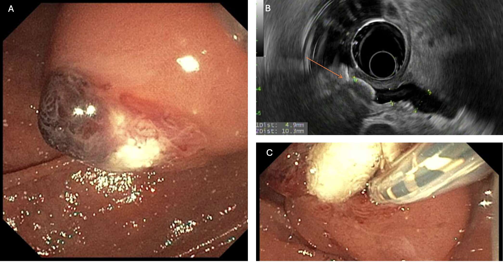
Figure: A. Impacted pancreatic duct stone at ampulla. B. Endoscopic ultrasound of pancreatic duct stone (orange arrow). C. Sphincterotomy and fragmentation of impacted stone.
Disclosures:
Omar Calderon indicated no relevant financial relationships.
Eric Vecchio indicated no relevant financial relationships.
Anurag Soni indicated no relevant financial relationships.
Deepak Gopal indicated no relevant financial relationships.
Omar Calderon, MD, Eric Vecchio, MD, Anurag Soni, MD, Deepak V. Gopal, MD, FACG. P3511 - Giant Impacted Pancreatic Duct Stone at the Ampulla of Vater, ACG 2024 Annual Scientific Meeting Abstracts. Philadelphia, PA: American College of Gastroenterology.
